. 2007 Dec;127(12):2807-17.
doi: 10.1038/sj.jid.5700970. Epub 2007 Jul 26.

Human bullous pemphigoid antigen 2 transgenic skin elicits specific IgG in wild-type mice

Affiliations

Human bullous pemphigoid antigen 2 transgenic skin elicits specific IgG in wild-type mice

Edit B Olasz et al. J Invest Dermatol. 2007 Dec.

Abstract

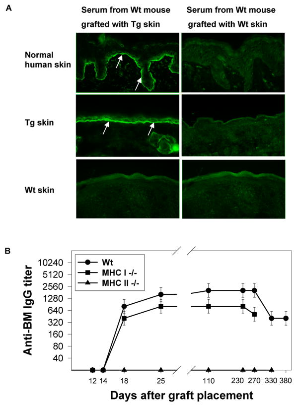
Bullous pemphigoid antigen 2 (BPAG2) is targeted by autoantibodies in patients with bullous pemphigoid (BP), and absent in patients with one type of epidermolysis bullosa (OMIM #226650). A keratin 14 promoter construct was used to produce transgenic (Tg) mice appropriately expressing human BPAG2 (hBPAG2) in murine epidermal basement membrane (BM). Grafts of Tg skin placed on gender-matched, syngeneic wild type (Wt) or major histocompatibility complex I (MHC I)-/- mice elicited IgG that bound human epidermal BM and BPAG2. Production of such IgG in grafted mice was prompt (detectable within 16+/-2 days), robust (titer > or = 1,280), durable (present > or = 380 days), and correlated with the involution and loss of Tg skin grafts. MHC II-/- mice grafted with Tg skin did not develop anti-hBPAG2 IgG or graft loss indicating that MHC II:CD4+ T cell interactions were crucial for these responses. Tg skin grafts on Wt mice developed neutrophil-rich infiltrates, dermal edema, subepidermal blisters, and deposits of immunoreactants in epidermal BM. This model shows fidelity to alterations seen in patients with BP, has relevance to immune responses that may arise in patients with epidermolysis bullosa following BPAG2 gene replacement, and can be used to identify interventions that may block production of IgG against proteins in epidermal BM.


Conflict of interest statement

Conflict of Interest: The authors state no conflict of interest.

Figures

Figure 1
Figure 1
The hBPAG2 cDNA transgene construct expressed protein of appropriate immunoreactivity and molecular weight in an immortalized GABEB keratinocyte line. a: An immortalized GABEB keratinocyte line transfected with the transgene construct showed bright, diffuse cytoplasmic staining for hBPAG2 in IF microscopy studies. b: In contrast, GABEB keratinocytes transfected with the same construct lacking hBPAG2 cDNA (i.e., cells transfected with a “mock” construct and subjected to selection in the same manner) showed no staining for hBPAG2. Nonimmune rabbit sera showed no reactivity to these cells (data not shown). c: Immunoblot studies of extracts from transgene- and mock-transfected GABEB keratinocytes identified hBPAG2 as a 180 kD polypeptide in the former (lane 4) but not the latter (lane 2). Immortalized GABEB keratinocytes transfected with the same hBPAG2 cDNA in an alternate vector (i.e., pcDNA3) served as an additional positive control (lane 3) as did extracts of normal HKs (lane 1). Nonimmune rabbit sera showed no reactivity to these extracts (data not shown). Ticks in the left margin correspond to 250 and 116 kD molecular weight markers; the arrow points to a 180 kD band corresponding to hBPAG2. d: Tg mice express hBPAG2 in epidermal BM. Cryosections of skin from man, a Tg mouse, and a Wt mouse (top, middle, and bottom panels, respectively) were studied by IF microscopy using rabbit antiserum developed against the carboxy terminus of hBPAG2 (Masunaga et al., 1997) (left panels) or normal rabbit serum (control, right panels). hBPAG2 was specifically expressed in the epidermal BMs of human and Tg skin (arrows, top and middle left panels); this antiserum showed no specific reactivity to the skin of a Wt mouse (bottom left panel). There was no cytosolic, suprabasal, or intradermal expression of hBPAG2 in any skin samples. Control IF microscopy studies using normal rabbit serum were negative (right panels).
Figure 2
Figure 2
hBPAG2 displayed normal ultrastructural localization in the epidermal BM of Tg skin. Post-embedding immunogold electron microscopy studies localized 5 nm gold particles staining hBPAG2 within the superior aspect of the lamina densa in human (a) and Tg (b) skin (arrows). This staining was consistent with that reported for this antiserum (Masunaga et al., 1997). Expression of hBPAG2 in Tg skin was somewhat less than that in human skin. There was no ectopic localization or aggregation of hBPAG2 in Tg skin. Anti-hBPAG2 antiserum showed no specific reactivity to the skin of a Wt mouse (c). HD, hemidesmosome; LL, lamina lucida; LD, lamina densa.
Figure 3
Figure 3
Tg skin grafts elicited anti-BM IgG in Wt and MHC I -/- mice. a. Tg skin grafts placed on Wt recipients elicited IgG that bound epidermal BMs in human and Tg skin (arrows, top and middle left panels), but not the skin of a Wt mouse (bottom left panel). Development of anti-BM IgG was unrelated to the grafting procedure (right panels). b: Longitudinal studies showed that Tg skin grafts placed on Wt (solid circles) and MHC I -/- (solid squares) recipients elicited high (640 to 2560) and durable (lasting ≥ 270 days) titers of anti-BM IgG that consistently appeared in the circulation of recipients 14 to 18 days after grafting. MHC II -/- mice (solid triangles) grafted with Tg skin did not develop anti-BM IgG. n=4/group in this longitudinal study that was representative of the aggregate work.
Figure 4
Figure 4
IgG in the sera of Wt mice grafted with Tg skin specifically bound hBPAG2. a. IgG in the sera of Wt mice grafted with Tg skin specifically bound hBPAG2 in extracts of cultured HKs as well as recombinant NC16A-GST (lanes 1 and 4, respectively). While preadsorption of immune sera with recombinant NC16A-GST completely removed all reactivity to this fusion protein (lane 5), immunoadsorbed immune sera still bound hBPAG2 (lane 2), indicating that epitopes other than NC16A are also targeted by such IgG. Normal mouse serum did not show any reactivity to hBPAG2 or NC16A-GST (lanes 3 and 6, respectively). Results were confirmed in three separate experiments. Markers in the left margins indicate Mr × 10-3. b. Immunoprecipitation studies of biosynthetically radiolabeled HK extracts showed that sera from Wt mice grafted with Tg skin (representative example, lane 1) contained IgG that bound BPAG2 but not BPAG1 – the former identified as a 180 kD protein that comigrated with BPAG2 immunoprecipitated by serum from a patient with BP (lane 3). Serum from a Wt mouse grafted with Wt skin (lane 2) as well as serum from a normal volunteer (lane 4) bound no specific HK proteins in these immunoprecipitation studies. Markers in the right margin indicate Mr × 10-3.
Figure 5
Figure 5
CD4+ T cells from Wt mice grafted with Tg skin demonstrated specific and dose-related proliferative responses to NC16A-GST in vitro. Ten days after receiving grafts of Tg skin, axillary and inguinal lymph nodes of Wt mice were harvested and processed to yield CD4+ T cells that were placed in culture and assessed for secondary proliferative responses to various concentrations of recombinant NC16A-GST (solid circles), recombinant GST alone (solid squares), or PBS (solid triangles). γ-irradiated splenocytes from naïve, syngeneic Wt mice were used as stimulator cells. CD4+ T cells from graft recipients proliferated specifically to NC16A-GST but not to GST alone or PBS. CD4+ T cells from Wt mice grafted with Wt skin did not show proliferative response to NC16A-GST, GST, or PBS (data not shown). n=10/group.
Figure 6
Figure 6
Wt and MHC I -/- mice lost Tg skin grafts with identical kinetics, while MHC II -/- mice retained grafts of Tg skin. Tail skin was harvested from Tg and Wt mice and grafted onto the opposite flanks of age- and gender-matched Wt (solid circles), MHC I -/- (solid squares), MHC II -/- (solid triangles), and “immune” Wt (open circles) recipients. Bandages were removed on day 7; thereafter, mice were examined daily for 30 days then weekly to assess graft viability and size. Skin grafts were graded as “lost” if their area became ≤ 20% of their original size (McFarland et al., 2003). These longitudinal studies demonstrated that grafts of Tg skin on Wt and MHC I -/- recipients displayed accelerated involution at days 15 to 23 (i.e., the approximate time circulating anti-hBPAG2 IgG became detectable in recipients), and complete loss by days 28 to 30. Tg skin placed on “immune” Wt recipients (i.e., mice previously grafted with Tg skin and displaying anti-hBPAG2 in their circulation) were typically lost within 15 days – a profile like that observed once anti-hBPAG2 IgG first appeared in Wt mice grafted with Tg skin. Tg skin grafts placed on MHC II -/- recipients retained their viability and size for their entire period of study. n=4/group in this longitudinal study that was representative of the aggregate work.
Figure 7
Figure 7
Tg skin grafts on Wt recipients developed neutrophil-rich leukocytic infiltrates and edema within the papillary dermis that eventuated in clefting and/or blister formation within the epidermal BM at sites where in situ deposits of IgG and C3 were localized. a: Tg and Wt skin was grafted onto the opposite flanks of age and gender-matched Wt, Tg, and MHC II -/- recipients; biopsies were obtained between 6 and 25 days after grafting for light and IF microscopy studies. All specific alterations (i.e., those unrelated to wounds at the base and margins of grafts) developed in Tg grafts on Wt recipients (shown here). Such alterations were evident as early as 17 days after grafting and consisted of neutrophil-rich infiltrates within the papillary dermis. b: Alterations in Tg skin grafts on Wt recipients progressed to cleft and/or subepidermal blister formation within 20 to 25 days (top panels). Direct IF microscopy revealed in situ deposits of murine IgG and C3 in epidermal BMs of such grafts (arrows, bottom panels). Immunoreactants were not identified in the epidermal BMs of recipient skin adjacent to, or at distance from, Tg grafts, or in control grafts of Wt skin placed on the opposite flanks of Wt (insets, bottom panels, dotted white lines outline epidermal BM), Tg, or MHC II -/- recipients. Scale bars, ∼25 μm.
Figure 8
Figure 8
Involution and loss of Tg skin grafts on CD4 -/- mice correlated with production of anti-BM IgG rather than exposure to CD4+ T cells (naïve or “immune”). Longitudinal studies showed that Tg skin did elicit anti-BM IgG in CD4 -/- recipients (solid circles), but that its production was delayed (day 44±5) in comparison to development of anti-BM IgG in Wt mice grafted with Tg skin (day 16±2). Accelerated graft loss in CD4 -/- recipients correlated with production of anti-BM IgG and progressed to completion within 12 to 15 days, a kinetic profile like that seen once specific IgG appeared in the circulation of Wt mice grafted with Tg skin. Adoptive transfer studies showed that involution and loss of Tg skin grafts on CD4 -/- recipients infused with naïve (solid squares) or “immune” (solid triangles) CD4+ T cells correlated better with the development of anti-BM IgG than exposure to CD4+ T cells. Arrows and brackets indicate the time points anti-BM IgG appeared in the circulation of CD4 -/- mice engrafted with Tg skin.

Similar articles

Cited by

References

    1. Darling TN, Bauer JW, Hintner H, Yancey KB. Generalized atrophic benign epidermolysis bullosa. Adv Dermatol. 1997a;13:87–119. - PubMed
    1. Darling TN, McGrath JA, Yee C, Gatalica B, Hametner R, Bauer JW, et al. Premature termination codons are present on both alleles of the bullous pemphigoid antigen 2/type XVII collagen gene in five Austrian families with generalized atrophic benign epidermolysis bullosa. J Invest Dermatol. 1997b;108:463–8. - PubMed
    1. Dumois JA, VanderVegt FP, Kopp JB, Marinos NJ, Rooney JF, Notkins AL. Transplantation of skin from human immunodeficiency virus type 1-transgenic mice to normal congenic mice results in graft rejection. J Infect Dis. 1995;172:232–4. - PubMed
    1. Ehst BD, Ingulli E, Jenkins MK. Development of a novel transgenic mouse for the study of interactions between CD4 and CD8 T cells during graft rejection. Am J Transplant. 2003;3:1355–62. - PubMed
    1. Franzke CW, Bruckner P, Bruckner-Tuderman L. Collagenous transmembrane proteins: recent insights into biology and pathology. J Biol Chem. 2005;280:4005–8. - PubMed

Publication types

MeSH terms