. 2007 Aug;117(8):2105-13.
doi: 10.1172/JCI32041.

Endothelial sulfonylurea receptor 1-regulated NC Ca-ATP channels mediate progressive hemorrhagic necrosis following spinal cord injury

Affiliations

Endothelial sulfonylurea receptor 1-regulated NC Ca-ATP channels mediate progressive hemorrhagic necrosis following spinal cord injury

J Marc Simard et al. J Clin Invest. 2007 Aug.

Abstract

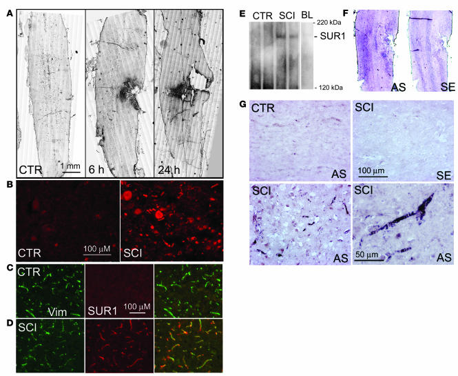
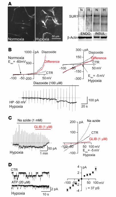
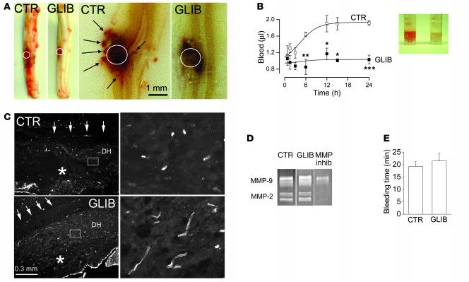
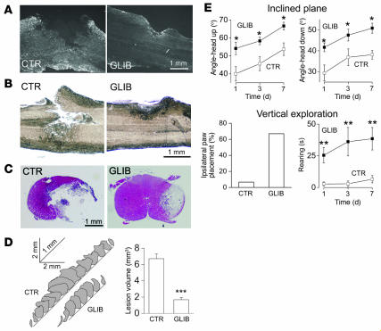
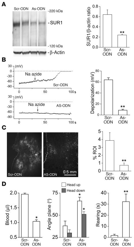
Acute spinal cord injury (SCI) causes progressive hemorrhagic necrosis (PHN), a poorly understood pathological process characterized by hemorrhage and necrosis that leads to devastating loss of spinal cord tissue, cystic cavitation of the cord, and debilitating neurological dysfunction. Using a rodent model of severe cervical SCI, we tested the hypothesis that sulfonylurea receptor 1-regulated (SUR1-regulated) Ca(2+)-activated, [ATP](i)-sensitive nonspecific cation (NC(Ca-ATP)) channels are involved in PHN. In control rats, SCI caused a progressively expansive lesion with fragmentation of capillaries, hemorrhage that doubled in volume over 12 hours, tissue necrosis, and severe neurological dysfunction. SUR1 expression was upregulated in capillaries and neurons surrounding necrotic lesions. Patch clamp of cultured endothelial cells exposed to hypoxia showed that upregulation of SUR1 was associated with expression of functional SUR1-regulated NC(Ca-ATP) channels. Following SCI, block of SUR1 by glibenclamide or repaglinide or suppression of Abcc8, which encodes for SUR1 by phosphorothioated antisense oligodeoxynucleotide essentially eliminated capillary fragmentation and progressive accumulation of blood, was associated with significant sparing of white matter tracts and a 3-fold reduction in lesion volume, and resulted in marked neurobehavioral functional improvement compared with controls. We conclude that SUR1-regulated NC(Ca-ATP) channels in capillary endothelium are critical to development of PHN and constitute a major target for therapy in SCI.


Figures

Figure 1
Figure 1. SUR1 is upregulated in SCI.
(A) Immunohistochemical localization of SUR1 in control rats (CTR) and at different times after SCI as indicated, with montages constructed from multiple individual images and positive labeling shown in black pseudocolor. (B) Magnified views of SUR1-immunolabeled sections taken from control and from the core (heavily labeled area in A at 6 hours). (C and D) Immunolabeling of capillaries with vimentin (Vim) and colabeling with SUR1 in control rats (C) and from the penumbra of SCI rats (tissue adjacent to the heavily labeled core in A, 6 hours) (D). (E) Western blots for SUR1 of spinal cord tissue from control rats (50 μg protein), from rats 6 hours after SCI (50 μg protein), and from an equivalent amount of blood (BL; 2 μl) as is present in the injured cord. Blots are representative of 5–6 control and SCI rats. (F and G) In situ hybridization for Abcc8 in control rats and in whole cords (F) or in the penumbra (G) 6 hours after SCI using antisense (AS) and sense (SE) as indicated. Immunohistochemistry and in situ hybridization images are representative of findings in 3–5 rats per group. Scale bars: 1 mm (A); 100 μM (BD and G, top panels and bottom left panel); 50 μM (G, bottom right panel).
Figure 2
Figure 2. The SUR1-regulated NCCa-ATP channel is upregulated in endothelial cells by hypoxia.
(A) Immunolabeling (scale bar: 50 μm) and Western blots for SUR1 in human aortic endothelial cells (ENDO) cultured under normoxic (N) or hypoxic (H) conditions as indicated, as well as Western blots for SUR1 of rat insulinoma RIN-m5F cells (INSUL) cultured under normoxic or hypoxic condition, with β-actin also shown. (B and C) Whole-cell currents during ramp pulses (4 per minute; holding potential [HP], –50 mV) or at the holding potential of –50 mV, before and after application of diazoxide (B) or Na azide (C), in endothelial cells exposed to normoxic or hypoxic conditions; the difference currents are also shown in red. Erev, reversal potential; GLIB, glibenclamide. Data are representative of 7–15 recordings from human aortic endothelial cells (B) or bEnd.3 cells (C) for each condition. (D) Single-channel recordings of inside-out patches with Cs+ as the principal cation, with channel openings inhibited by ATP on the cytoplasmic side; channel amplitude at various potentials indicated a slope conductance of 37 pS (data from 7 patches) from human brain microvascular endothelial cells. Error bars indicate SEM.
Figure 3
Figure 3. Block of SUR1 reduces hemorrhage after SCI.
(A) Whole cords and longitudinal sections of cords 24 hours after SCI, from vehicle-treated control and glibenclamide-treated rats. White circles indicate site of impact; arrows denote petechial hemorrhages. (B) Cord homogenates in test tubes at 24 hours (inset) and spectrophotometric measurements of blood in cord homogenates at various times after SCI from vehicle-treated (n = 66) and glibenclamide-treated (n = 62) rats. *P < 0.05, **P < 0.01, ***P < 0.001 versus control. (C) Cord sections immunolabeled for vimentin to show capillaries from SCI rats treated with vehicle or glibenclamide; arrows indicate the central canal. Right panels are higher-magnification images of boxed areas in left panels. Images are representative of findings in 6 rats per group. Asterisks indicate lesion core at impact site. DH, dorsal horn. (D) Zymography of recombinant MMP-2 and MMP-9 performed under control conditions, in the presence of glibenclamide (10 μM), and in the presence of MMP inhibitor II (MMP inhib; 300 nM). (E) Bleeding times in uninjured rats infused with vehicle or glibenclamide (n = 3 per group). Error bars indicate SEM. Scale bars: 1 mm (A); 0.3 mm (C, left panels). Original magnification, ×40 (C, right panels).
Figure 4
Figure 4. Blocking SUR1 reduces lesion size and improves neurobehavioral function after SCI.
(AC) Cord sections immunolabeled for glial fibrillary acidic protein (A) or stained with eriochrome cyanine R (B) or H&E (C), 1 day (A and B) or 7 days (C) after SCI, from vehicle-treated and glibenclamide-treated rats. Images are representative of findings in 3 rats per group. Scale bars: 1 mm. (D) Cascaded outlines of lesion areas in serial sections 250 μm apart, 7 days after SCI, as well as lesion volumes from vehicle-treated and glibenclamide-treated rats (n = 4–6 per group; excludes 2 control rats that died). (E) Performance on inclined plane (head up and head down), ipsilateral paw placement, and rearing in the same vehicle-treated and glibenclamide-treated rats as in D. Paw placement was measured 1 day after SCI. Error bars indicate SEM. *P < 0.05, **P < 0.01, ***P < 0.001 versus control.
Figure 5
Figure 5. Gene suppression of SUR1 blocks expression of functional NCCa-ATP channels and improves outcome in SCI.
(A) Western blots for SUR1 in gliotic capsule from rats with infusion of scrambled ODN (Scr-ODN) or antisense ODN (AS-ODN) directly into the brain injury site for 10–12 days prior to tissue harvest. Also shown is densitometric analysis of Western blots from the same groups of rats (n = 3 per group). (B) Membrane potential of astrocytes from gliotic capsules of the same groups of rats as in A during application of Na azide to deplete ATP. Mean depolarization of 3 cells per group is shown. (C) Cord sections immunolabeled for SUR1, 1 day after SCI, from rats treated with i.v. infusion of scrambled ODN or antisense ODN. Scale bar: 0.5 mm. Also shown is quantitative immunofluorescence for the same groups of rats (n = 3 per group). ROI, region of interest. (D) Blood in cord homogenates, performance on angled plane, and rearing, 1 day after SCI, for rats treated with i.v. infusion of scrambled ODN or antisense ODN. Error bars indicate SEM. *P < 0.05, **P < 0.01 versus scrambled ODN.

References

    1. Hayes K.C., Kakulas B.A. Neuropathology of human spinal cord injury sustained in sports-related activities. J. Neurotrauma. 1997;14:235–248. - PubMed
    1. Tator C.H., Fehlings M.G. Review of the secondary injury theory of acute spinal cord trauma with emphasis on vascular mechanisms. J. Neurosurg. 1991;75:15–26. - PubMed
    1. Kwon B.K., Tetzlaff W., Grauer J.N., Beiner J., Vaccaro A.R. Pathophysiology and pharmacologic treatment of acute spinal cord injury. Spine J. 2004;4:451–464. - PubMed
    1. Bilgen M., Abbe R., Liu S.J., Narayana P.A. Spatial and temporal evolution of hemorrhage in the hyperacute phase of experimental spinal cord injury: in vivo magnetic resonance imaging. Magn. Reson. Med. 2000;43:594–600. - PubMed
    1. Nelson E., Gertz S.D., Rennels M.L., Ducker T.B., Blaumanis O.R. Spinal cord injury. The role of vascular damage in the pathogenesis of central hemorrhagic necrosis. Arch. Neurol. 1977;34:332–333. - PubMed

Publication types

MeSH terms