Review
. 2007 Sep;102(5):369-92.
doi: 10.1007/s00395-007-0666-z. Epub 2007 Jul 27.

Excitation-contraction coupling and mitochondrial energetics

Affiliations
Review

Excitation-contraction coupling and mitochondrial energetics

Christoph Maack et al. Basic Res Cardiol. 2007 Sep.

Abstract

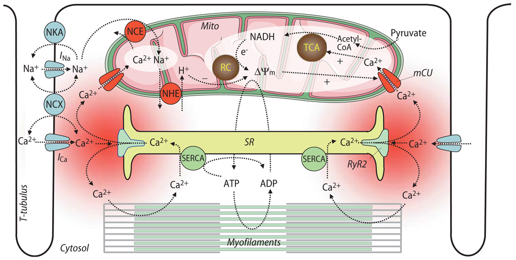
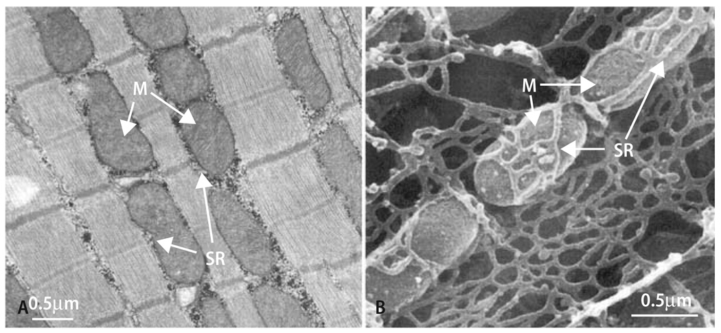
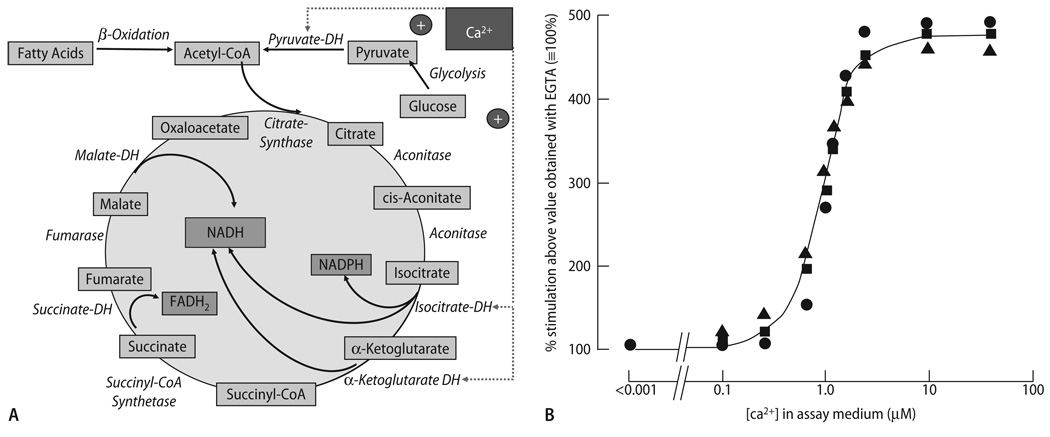
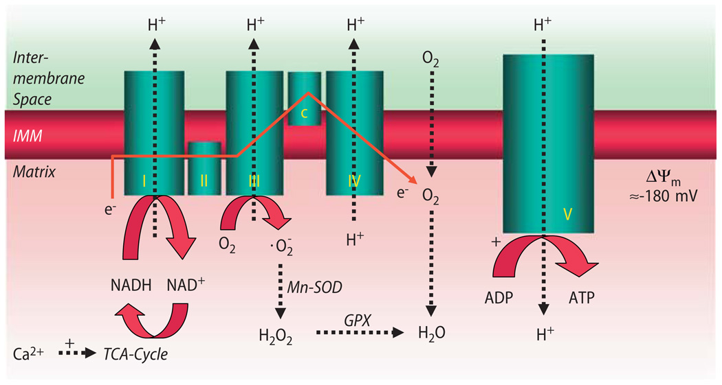
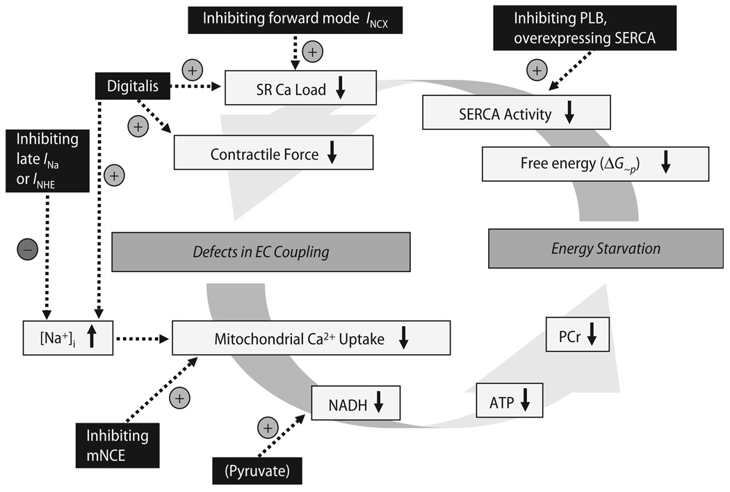
Cardiac excitation-contraction (EC) coupling consumes vast amounts of cellular energy, most of which is produced in mitochondria by oxidative phosphorylation. In order to adapt the constantly varying workload of the heart to energy supply, tight coupling mechanisms are essential to maintain cellular pools of ATP, phosphocreatine and NADH. To our current knowledge, the most important regulators of oxidative phosphorylation are ADP, Pi, and Ca2+. However, the kinetics of mitochondrial Ca2+-uptake during EC coupling are currently a matter of intense debate. Recent experimental findings suggest the existence of a mitochondrial Ca2+ microdomain in cardiac myocytes, justified by the close proximity of mitochondria to the sites of cellular Ca2+ release, i. e., the ryanodine receptors of the sarcoplasmic reticulum. Such a Ca2+ microdomain could explain seemingly controversial results on mitochondrial Ca2+ uptake kinetics in isolated mitochondria versus whole cardiac myocytes. Another important consideration is that rapid mitochondrial Ca2+ uptake facilitated by microdomains may shape cytosolic Ca2+ signals in cardiac myocytes and have an impact on energy supply and demand matching. Defects in EC coupling in chronic heart failure may adversely affect mitochondrial Ca2+ uptake and energetics, initiating a vicious cycle of contractile dysfunction and energy depletion. Future therapeutic approaches in the treatment of heart failure could be aimed at interrupting this vicious cycle.


Figures

Fig. 1
Fig. 1
Overview on processes of excitation-contraction coupling and mitochondrial energetics. SR sarcoplasmic reticulum; SERCA SR Ca2+ ATPase; Mito mitochondria; TCA tricarboxylic acid cycle; RC respiratory chain; Δψm mitochondrial membrane potential; NCE mitochondrial Na+/Ca2+-exchanger; NHE mitochondrial Na+/H+ -exchanger; NKA sarcolemmal Na+/K+-ATPase; NCX sarcolemmal Na+/Ca2+-exchanger; RyR2 ryanodine receptor type 2; mCU mitochondrial Ca2+-uniporter; INa and ICa, currents of voltage-gated Na+- or Ca2+-channels, respectively
Fig. 2
Fig. 2
Longitudinal (A) or scanning (B) electron micrographs of cardiac myocytes. M mitochondria; SR sarcoplasmic reticulum. With permission from Territo et al. [194] and Yoshikane et al. [220], respectively
Fig. 3
Fig. 3
(A) Enzymatic reactions of the tricarboxylic acid (TCA) cycle, and their regulation by Ca2+. DH dehydrogenase. (B) Stimulation of pyruvate-dehydrogenase phosphatase (PDH-PPase), NAD-isocitrate dehydrogeNase (NAD-ICDH) and α-ketoglutarate dehydrogenase (α-KGDH) enzymatic activity by Ca2+ in extracts of mitochondria. Activity in the presence of EGTA was ∼ 10–20% of maximal velocity (Vmax), respectively. Reproduced from Denton & McCormack [54] with permission
Fig. 4
Fig. 4
Processes of oxidative phosphorylation at the mitochondrial respiratory chain. I–V complexes I–V of the respiratory chain. Complex V F1/F0-ATPase; c cytochrome c. Electrons (e) donated by NADH to complex I (and by succinate to complex II; not shown) elicit sequential redox reactions at complexes I–IV, promoting H+-translocation from the matrix to the intermembrane space across the inner mitochondrial membrane (IMM). This creates a proton gradient (ΔpH), which together with the electrochemical gradient (Δψm) constitutes the driving force for protons to flow back into the matrix space via F1/F0-ATPase, promoting ATP-production from ADP. At complex III (and I; not shown), the superoxide anion radical is produced already under physiological conditions. It is dismutated to hydrogen peroxide (H2O2) by Mn2+-dependent superoxide-dismutase (Mn-SOD), which is eliminated to H2O by glutathione-peroxidase (GPX). While Ca2+ activates (+) TCA-cycle dehydrogenases (but also complex V of the respiratory chain; not shown), ADP regulates respiration by increasing complex V activity (+)
Fig. 5
Fig. 5
[Ca2+]c and [Ca2+]m during a single contraction in the presence and absence of norepinephrine (NE): myocyte loaded with indo-1/salt (A); myocyte loaded with indo-1/AM and quenched with Mn2+ (B). A and B each show results from a single cell, stimulated at steady-state 0.2 Hz in presence and absence of 1 µM NE. It is seen that there is no response of [Ca2+]m to a single electrical stimulation. (C) Kinetics of rise and fall of [Ca2+]m in response to changed pacing rate. Top: [Ca2+]m for a single myocyte, superfused with medium containing 1 µM NE; bottom: mechanical response of cell to electrical stimulation. At 1st arrow, stimulation frequency was increased from 0 to 4 Hz; at 2nd arrow, cell begins to contract in synchrony with 4 Hz stimulation; at 3rd arrow, pacing was discontinued. (D) Relation between [Ca2+]m and mean [Ca2+]c during Na+-replacement experiments with slow cytosolic and mitochondrial Ca2+ uptake. Reproduced with permission from Miyata et al. [137]
Fig. 6
Fig. 6
(A-D) Representative experiment determining [Ca2+]c and [Ca2+]m in the same cell. [Ca2+]c transients were elicited at 1 Hz in the absence and presence of isoproterenol (Iso, 1–100 nmol/L; (A), (C)). The [Ca2+] transients in (B) and (D) were recorded at the indicated time points of the protocol (arrows in (A), (C)). (E) and (F) Cytosolic (E) and mitochondrial Ca2+-transients (F) in the absence and presence of Ru360 (10 nmol/L)
Fig. 7
Fig. 7
Full activation of mitochondrial Ca2+ uptake during RyR-mediated Ca2+ release. (A) Ca2+- and caffeine induced [Ca2+]m and Δψm responses were measured in permeabilized myotubes using rhod-2 and TMRM, respectively. Uncoupler (1 µM CCCP and 2.5 µg/ml oligomycin) was added as indicated. (B) The rate of mitochondrial Ca2+ uptake was measured at varying [Ca2+] obtained by the addition of CaCl2 in adherent rhod-2 loaded permeabilized cells. The added CaCl2 concentration values are indicated with arrows below the x axis, and the effective [Ca2+]c were calculated. To prevent Ca2+-induced Ca2+-release via RyR2, the cells were preincubated with thapsigargin to inhibit the SR Ca2+ ATPase and hence to deplete the SR prior to Ca2+ addition. Reproduced from Szalai et al. [191] with permission
Fig. 8
Fig. 8
Computational modeling of [Ca2+]m, assuming a microdomain (MD) in which mitochondria are exposed to [Ca2+] pulses of 10–20 µmol/L for 50 ms (D; representative pulse taken from t = 215 s in A). Starting from steady-state conditions with [Ca2+]MD oscillating from 0.1 to 10 µmol/L at 1 Hz, an increase of amplitude (A, from 10 to 20 µmol/L), frequency (B, from 1 to 2 Hz) or both (C) of [Ca2+]MD transients were simulated. Reproduced from Maack et al. [117] with permission
Fig. 9
Fig. 9
Time-dependent behavior of [Ca2+]m and NADH after changes in workload. (A) Experimental data showing the responses of NADH (upper panel) and [Ca2+]m (lower panel) to an increase in stimulation frequency from 0.5 to 2 Hz in a rat cardiac trabecula. (B) NADH (upper panel) and [Ca2+]m (lower panel) in response to an increase of stimulation frequency from 0 to 4 Hz in the presence of isoproterenol (100 nM). (C) Computational modeling of NADH and [Ca2+]m by integrating the kinetics of EC coupling, mitochondrial Ca2+ transporters and energetics in response to a change in stimulation frequency from 0.25 Hz to 2 Hz (for parameters, see [46]). I, undershoot; II, recovery, and III, overshoot of NADH. Reproduced from Brandes and Bers (A; [26]), Maack et al. (B; [117]) and Cortassa et al. (C; [46]) with permission
Fig. 10
Fig. 10
Effect of changing the actomyosin- (AM-) ATPase and mCU on the computational model (described by Cortassa et al. [46]; compare Fig. 9C) on energetic behavior after changes in workload. (A) The behavior of NADH with the maximal rate of ATP hydrolysis by the AM-ATPase being decreased to onehalf (solid trace; Vmax/AM = 3.6·10−3 mM ms−1) of the control value (shaded trace). (B) The profile of average ATPi for changes in workload under control conditions (shaded trace) or with decreased AM-ATPase activity (solid trace). (C) The NADH profile with the maximal rate of the mCU decreased to 1/10th (solid trace; Vmax/mCU = 2.75·10−3 mM ms−1) of the control value (shaded trace). (D) The profile of [Ca2+]m accumulation for the low mCU condition described in (C) (solid trace) as compared to the control (shaded trace). Reproduced from Cortassa et al. [46] with permission
Fig. 11
Fig. 11
Stimulation of α-ketoglutarate oxidation by Ca2+ in the absence and presence of Na+ (15 mM) and the mCU-inhibitor ruthenium red (RR). Reproduced from Denton et al. [55] with permission
Fig. 12
Fig. 12
[Ca2+]c (A), [Ca2+]m (B) and reduced NADH (C) in guinea-pig cardiac myocytes paced at 3 Hz and exposure to 10 and 100 nM isoproterenol, with either 5 or 15 mM [Na+] in the pipet. Reproduced from Maack et al. [117] with permission
Fig. 13
Fig. 13
Hypothetical vicious cycle (and potential targets for therapeutic interventions) of defects in EC coupling and energy depletion in chronic heart failure. PCr phosphocreatine; PLB phospholamban; SERCA SR Ca2+-ATPase

Similar articles

Cited by

References

    1. Abozguia K, Clarke K, Lee L, Frenneaux M. Modification of myocardial substrate use as a therapy for heart failure. Nat Clin Pract Cardiovasc Med. 2006;3:490–498. - PubMed
    1. Aker S, Snabaitis AK, Konietzka I, Van De Sand A, Bongler K, Avkiran M, Heusch G, Schulz R. Inhibition of the Na+/H+ exchanger attenuates the deterioration of ventricular function during pacing-induced heart failure in rabbits. Cardiovasc Res. 2004;63:273–282. - PubMed
    1. Armoundas AA, Hobai IA, Tomaselli GF, Winslow RL, O’Rourke B. Role of sodium-calcium exchanger in modulating the action potential of ventricular myocytes from normal and failing hearts. Circ Res. 2003;93:46–53. - PMC - PubMed
    1. Auffermann W, Wu ST, Parmley WW, Wikman-Coffelt J. Glycolysis in heart failure: a 31P-NMR and surface fluorometry study. Basic Res Cardiol. 1990;85:342–357. - PubMed
    1. Baartscheer A, Schumacher CA, van Borren MM, Belterman CN, Coronel R, Fiolet JW. Increased Na+/H+-exchange activity is the cause of increased [Na+]i and underlies disturbed calcium handling in the rabbit pressure and volume overload heart failure model. Cardiovasc Res. 2003;57:1015–1024. - PubMed

Publication types