Finite element analysis of microelectrotension of cell membranes
- PMID: 17657517
- PMCID: PMC3251963
- DOI: 10.1007/s10237-007-0093-y
Finite element analysis of microelectrotension of cell membranes
Abstract
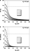
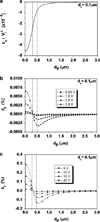
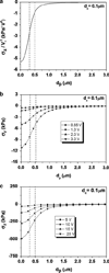
Electric fields can be focused by micropipette-based electrodes to induce stresses on cell membranes leading to tension and poration. To date, however, these membrane stress distributions have not been quantified. In this study, we determine membrane tension, stress, and strain distributions in the vicinity of a microelectrode using finite element analysis of a multiscale electro-mechanical model of pipette, media, membrane, actin cortex, and cytoplasm. Electric field forces are coupled to membranes using the Maxwell stress tensor and membrane electrocompression theory. Results suggest that micropipette electrodes provide a new non-contact method to deliver physiological stresses directly to membranes in a focused and controlled manner, thus providing the quantitative foundation for micreoelectrotension, a new technique for membrane mechanobiology.
Figures


Similar articles
-
Estimating the sensitivity of mechanosensitive ion channels to membrane strain and tension.Biophys J. 2004 Oct;87(4):2870-84. doi: 10.1529/biophysj.104.040436. Biophys J. 2004. PMID: 15454477 Free PMC article.
-
Finite element analysis of imposing femtonewton forces with micropipette aspiration.Ann Biomed Eng. 2002 Apr;30(4):546-54. doi: 10.1114/1.1476017. Ann Biomed Eng. 2002. PMID: 12086005
-
A finite-element model of the mechanical effects of implantable microelectrodes in the cerebral cortex.J Neural Eng. 2005 Dec;2(4):103-13. doi: 10.1088/1741-2560/2/4/006. Epub 2005 Oct 11. J Neural Eng. 2005. PMID: 16317234
-
Electric field-induced effects on neuronal cell biology accompanying dielectrophoretic trapping.Adv Anat Embryol Cell Biol. 2003;173:III-IX, 1-77. doi: 10.1007/978-3-642-55469-8. Adv Anat Embryol Cell Biol. 2003. PMID: 12901336 Review.
-
Caveolae - mechanosensitive membrane invaginations linked to actin filaments.J Cell Sci. 2015 Aug 1;128(15):2747-58. doi: 10.1242/jcs.153940. Epub 2015 Jul 9. J Cell Sci. 2015. PMID: 26159735 Review.
Cited by
-
Vesicle biomechanics in a time-varying magnetic field.BMC Biophys. 2015 Jan 21;8(1):2. doi: 10.1186/s13628-014-0016-0. eCollection 2015. BMC Biophys. 2015. PMID: 25649322 Free PMC article.
-
Multiscale modelling of the extracellular matrix.Matrix Biol Plus. 2021 Dec 14;13:100096. doi: 10.1016/j.mbplus.2021.100096. eCollection 2022 Feb. Matrix Biol Plus. 2021. PMID: 35072037 Free PMC article.
-
Mechanic stress generated by a time-varying electromagnetic field on bone surface.Med Biol Eng Comput. 2018 Oct;56(10):1793-1805. doi: 10.1007/s11517-018-1814-3. Epub 2018 Mar 19. Med Biol Eng Comput. 2018. PMID: 29556951
-
Contact-free scanning and imaging with the scanning ion conductance microscope.Anal Chem. 2014 Mar 4;86(5):2353-60. doi: 10.1021/ac402748j. Epub 2014 Feb 12. Anal Chem. 2014. PMID: 24521282 Free PMC article.
References
-
- Ashkin A, Dziedzic JM, Yamane T. Optical trapping and manipulation of single cells using infrared laser beams. Nature. 1987;330:769–771. - PubMed
Publication types
MeSH terms
Substances
Grants and funding
LinkOut - more resources
Full Text Sources
