

Alpha spectral power and coherence in the patients with mild cognitive impairment during a three-level working memory task
- PMID: 17657862
- PMCID: PMC1934955
- DOI: 10.1631/jzus.2007.B0584
Alpha spectral power and coherence in the patients with mild cognitive impairment during a three-level working memory task
Abstract
Objective: The functional relationship between calculated alpha band spectral power and inter-/intra-hemispheric coherence during a three-level working memory task of patients with mild cognitive impairment (MCI) was investigated.
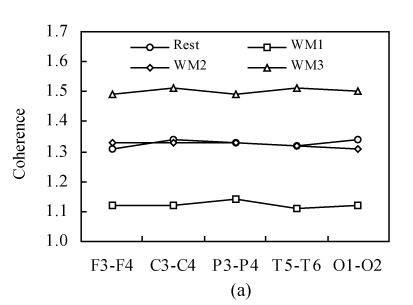
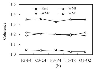
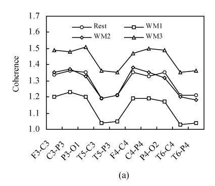
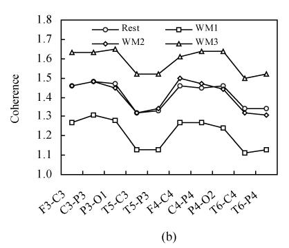
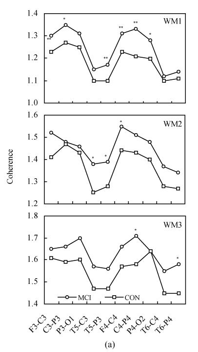
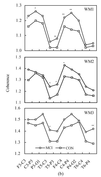
Methods: Subjects included 35 MCI patients according to the DSM-IV criteria (mean age: 62.3, SD: 6.5) and 34 healthy controls (mean age: 57.4, SD: 4.0) were selected from the community at large. All subjects performed a simple calculation and recall task with three levels of working memory load while electroencephalograph (EEG) signal was recorded. The spectral EEG power was computed over alpha1 (8.0-10.0 Hz) and alpha2 (10.5-13.0 Hz) frequency bands and was compared between rest stage and working memory processing stage by two-way ANOVA. Post hoc testing analyzed the differences between each two levels of working memory load during task processing. The inter-hemisphere EEG coherence of frontal (F3-F4), central (C3-C4), parietal (P3-P4), temporal (T5-T6) as well as occipital (O1-O2) was compared between MCI patients and normal controls. The EEG signals from F3-C3, F4-C4, C3-P3, C4-P4, P3-O1, P4-O2, T5-C3, T6-C4, T5-P3 and T6-P4 electrode pairs resulted from the intra-hemispheric action for alpha1 and alpha2 frequency bands.
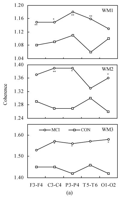
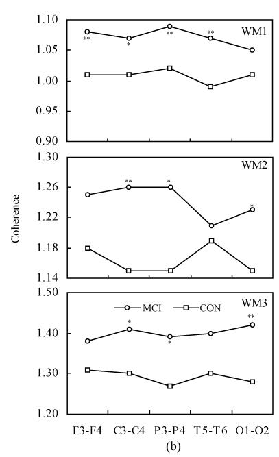
Result: There was significantly higher EEG power from MCI patients than from normal controls both at rest and during working memory processing. Significant differences existed between rest condition and three-level working memory tasks (P<0.001). The inter- and intra-hemispheric coherence during working memory tasks showed a "drop to rise" tendency compared to that at rest condition. There was significantly higher coherence in MCI patients than in the controls. When task difficulties increased, the cortical connectivity of intra-hemispheric diminished while the inter-hemispheric connectivity dominantly maintained the cognitive processing in MCI patients.
Conclusion: The results of the present study indicate that the alpha frequency band may be the characteristic band in distinguishing MCI patients from normal controls during working memory tasks. MCI patients exhibit greater inter-hemispheric connectivity than intra-hemispheric connectivity when memory demands increase. MCI patients mobilize a compensatory mechanism to maintain the processing effectiveness while the processing efficiency is reduced.
Figures
Similar articles
-
Study on EEG power and coherence in patients with mild cognitive impairment during working memory task.J Zhejiang Univ Sci B. 2005 Dec;6(12):1213-9. doi: 10.1631/jzus.2005.B1213. J Zhejiang Univ Sci B. 2005. PMID: 16358382 Free PMC article. Clinical Trial.
-
Inter- and intra-hemispheric EEG coherence in patients with mild cognitive impairment at rest and during working memory task.J Zhejiang Univ Sci B. 2006 May;7(5):357-64. doi: 10.1631/jzus.2006.B0357. J Zhejiang Univ Sci B. 2006. PMID: 16615165 Free PMC article.
-
EEG coherence characteristics at rest and during a three-level working memory task in normal aging and mild cognitive impairment.Med Sci Monit. 2008 Oct;14(10):CR515-23. Med Sci Monit. 2008. PMID: 18830191
-
Mild Cognitive Impairment: Structural, Metabolical, and Neurophysiological Evidence of a Novel EEG Biomarker.Front Neurol. 2015 Jul 6;6:152. doi: 10.3389/fneur.2015.00152. eCollection 2015. Front Neurol. 2015. PMID: 26217299 Free PMC article. Review.
-
Prefrontal cortex asymmetry and psychological responses to exercise: A systematic review.Physiol Behav. 2019 Sep 1;208:112580. doi: 10.1016/j.physbeh.2019.112580. Epub 2019 Jun 17. Physiol Behav. 2019. PMID: 31220517
Cited by
-
Quantitative EEG Markers in Mild Cognitive Impairment: Degenerative versus Vascular Brain Impairment.Int J Alzheimers Dis. 2012;2012:917537. doi: 10.1155/2012/917537. Epub 2012 Jul 26. Int J Alzheimers Dis. 2012. PMID: 22900229 Free PMC article.
-
Anatomical Substrate and Scalp EEG Markers are Correlated in Subjects with Cognitive Impairment and Alzheimer's Disease.Front Psychiatry. 2011 Jan 4;1:152. doi: 10.3389/fpsyt.2010.00152. eCollection 2011. Front Psychiatry. 2011. PMID: 21423459 Free PMC article.
-
Functional and effective EEG connectivity patterns in Alzheimer's disease and mild cognitive impairment: a systematic review.Front Aging Neurosci. 2025 Feb 12;17:1496235. doi: 10.3389/fnagi.2025.1496235. eCollection 2025. Front Aging Neurosci. 2025. PMID: 40013094 Free PMC article.
-
Coherence in event-related EEG oscillations in patients with Alzheimer's disease dementia and amnestic mild cognitive impairment.Cogn Neurodyn. 2023 Dec;17(6):1621-1635. doi: 10.1007/s11571-022-09920-0. Epub 2022 Dec 17. Cogn Neurodyn. 2023. PMID: 37974589 Free PMC article.
-
Synchronization during an internally directed cognitive state in healthy aging and mild cognitive impairment: a MEG study.Age (Dordr). 2014 Jun;36(3):9643. doi: 10.1007/s11357-014-9643-2. Age (Dordr). 2014. PMID: 24658709 Free PMC article.
References
-
- American Psychiatric Association. Diagnostic and Statistical Manual of Mental Disorders. 4th Ed. Washington, DC: APA Press; 1994. (DSM-IV)
-
- deToledo-Morrell L, Evers S, Hoeppner TJ, Morrell F, Garron DC, Fox JH. A ‘stress’ test for memory dysfunction. Electrophysiogical manifestation of early Alzheimer’s disease. Arch Neurol. 1991;48(6):605–609. - PubMed
Publication types
MeSH terms
LinkOut - more resources
Full Text Sources
Medical
Miscellaneous
