

Catecholaminergic neuronal loss in locus coeruleus of aged female dtg APP/PS1 mice
- PMID: 17658239
- PMCID: PMC5483173
- DOI: 10.1016/j.jchemneu.2007.05.008
Catecholaminergic neuronal loss in locus coeruleus of aged female dtg APP/PS1 mice
Abstract
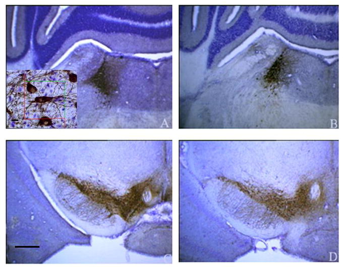
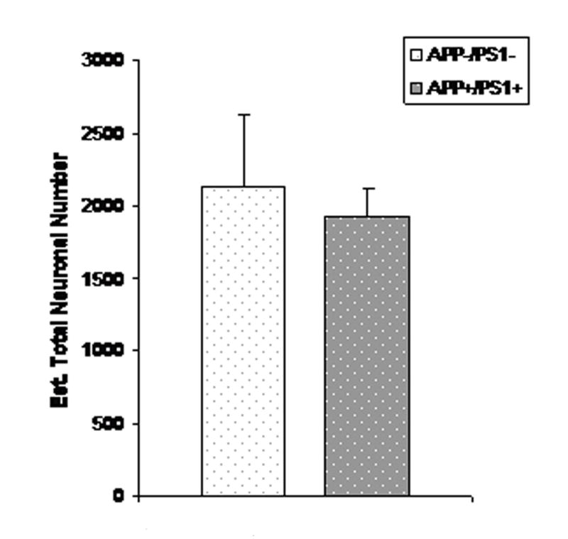
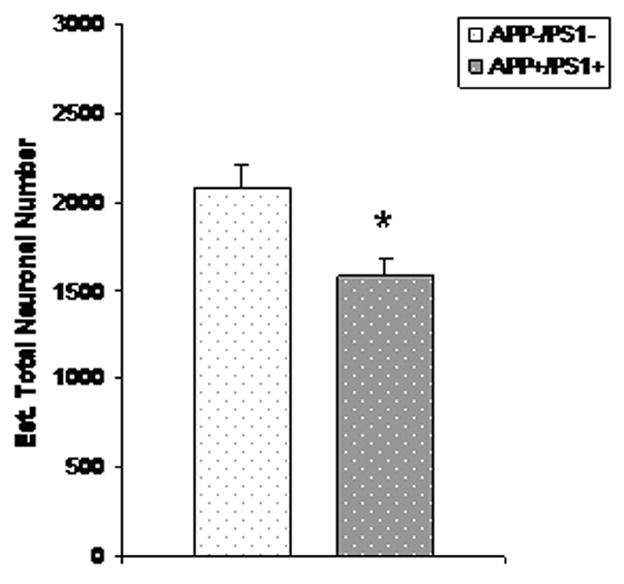
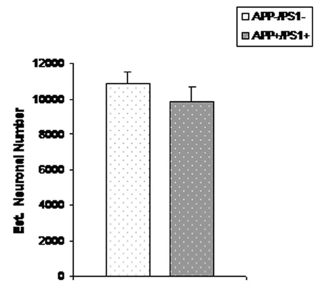
Alzheimer's disease (AD) is the most common type of dementia afflicting the elderly. In addition to the presence of cortical senile plaques and neurofibrillary tangles, AD is characterized at autopsy by extensive degeneration of brainstem locus coeruleus (LC) neurons that provide noradrenergic innervation to cortical neuropil, together with relative stability of dopaminergic neuron number in substantia nigra (SN) and ventral tegmental area (VTA). The present study used design-based stereological methods to assess catecholaminergic neuronal loss in brains of double transgenic female mice that co-express two human mutations associated with familial AD, amyloid precursor protein (APP(swe)) and presenilin-1 (PS1(DeltaE9)). Mice were analyzed at two age groups, 3-6 months and 16-23 months, when deposition of AD-type beta-amyloid (Abeta) plaques occurs in cortical brain regions. Blocks of brain tissue containing the noradrenergic LC nucleus and two nuclei of dopaminergic neurons, the SN and VTA, were sectioned and sampled in a systematic-random manner and immunostained for tyrosine hydroxylase (TH), a specific marker for catecholaminergic neurons. Using the optical fractionator method we found a 24% reduction in the total number of TH-positive neurons in LC with no changes in SN-VTA of aged dtg APP/PS1 mice compared with non-transgenic controls. No significant differences were observed in numbers of TH-positive neurons in LC or SN-VTA in brains of young female dtg APP/PS1 mice compared to their age-matched controls. The findings of selective neurodegeneration of LC neurons in the brains of aged female dtg APP/PS1 mice mimic the neuropathology in the brains of AD patients at autopsy. These findings support the use of murine models of Abeta deposition to develop novel strategies for the therapeutic management of patients afflicted with AD.
Figures
References
-
- Alzheimer A. Ueber eine eigenartige Erkrankung der Hirnrinde. Allgemeine Zeitschrift fur Psychiatrie. 1907;64:146–148.
-
- Aletrino MA, Vogels OJ, Van Domburg PH, Ten Donkelaar HJ. Cell loss in the nucleus raphes dorsalis in Alzheimer’s disease. Neurobiol Aging. 1992;13(4):461–8. - PubMed
-
- Astony-Jones G, Rajkowski J, Cohen J. Role of locus coeruleus in attention and behavioral flexibility. Biol Psychiatry. 1999;46:1309–1320. - PubMed
-
- Borchelt DR, Ratovitski T, van Lare J, Lee MK, Gonzales V, Jenkins NA, Copeland NG, Price DL, Sisodia SS. Accelerated amyloid deposition in the brains of transgenic mice coexpressing mutant presenilin 1 and amyloid precursor proteins. Neuron. 1997;19:939–45. - PubMed
-
- Busch C, Bohl J, Ohm TG. Spatial, temporal and numeric analysis of Alzheimer changes in the nucleus coeruleus. Neurobiol Aging. 1997;18:401–406. - PubMed
Publication types
MeSH terms
Substances
Grants and funding
LinkOut - more resources
Full Text Sources
Medical
Molecular Biology Databases
