. 2007 Aug 7;17(15):1334-9.
doi: 10.1016/j.cub.2007.06.060. Epub 2007 Jul 19.

UNC-1 regulates gap junctions important to locomotion in C. elegans

Affiliations

UNC-1 regulates gap junctions important to locomotion in C. elegans

Bojun Chen et al. Curr Biol. .

Abstract

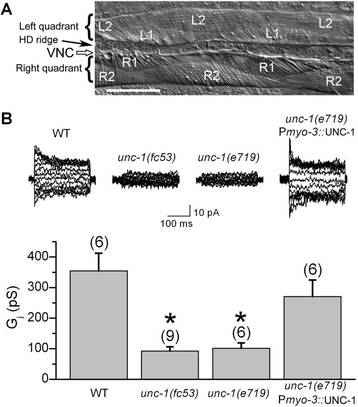
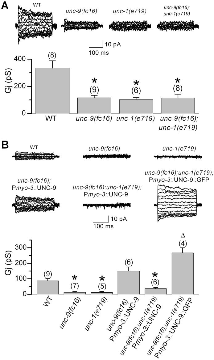
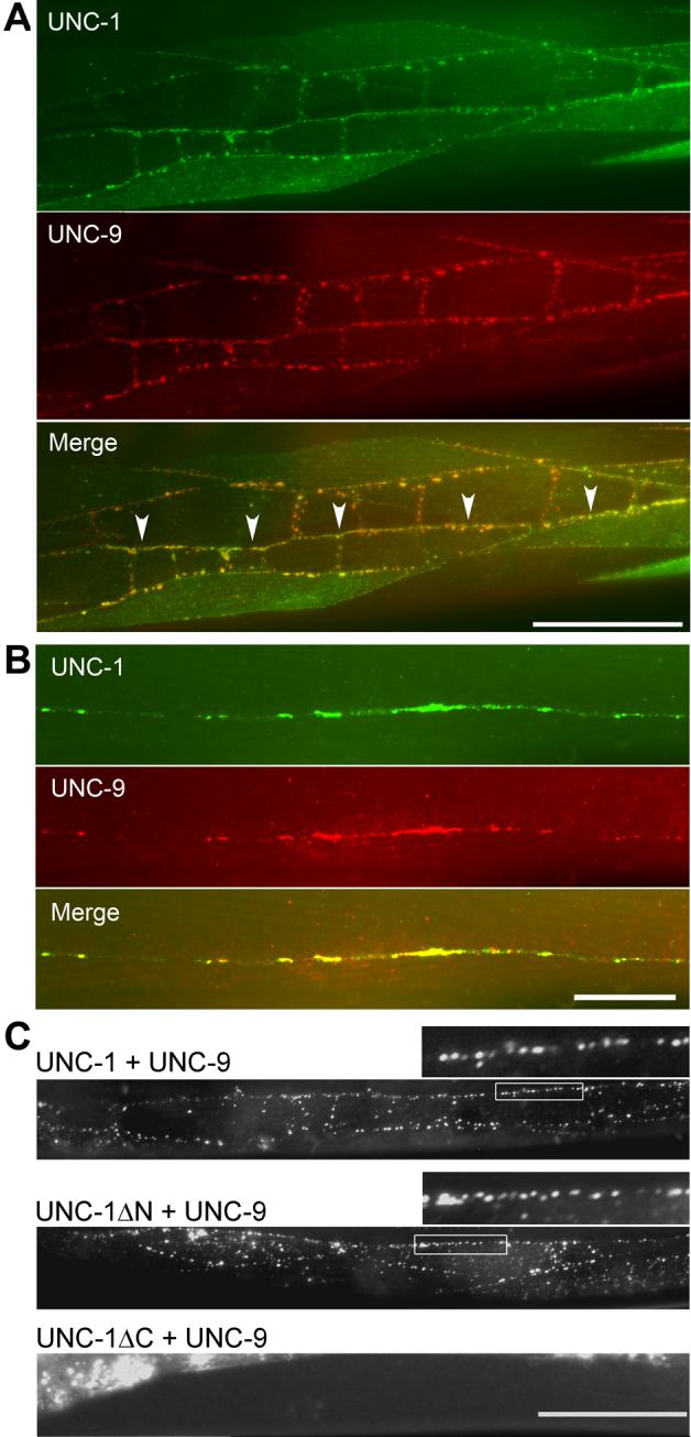
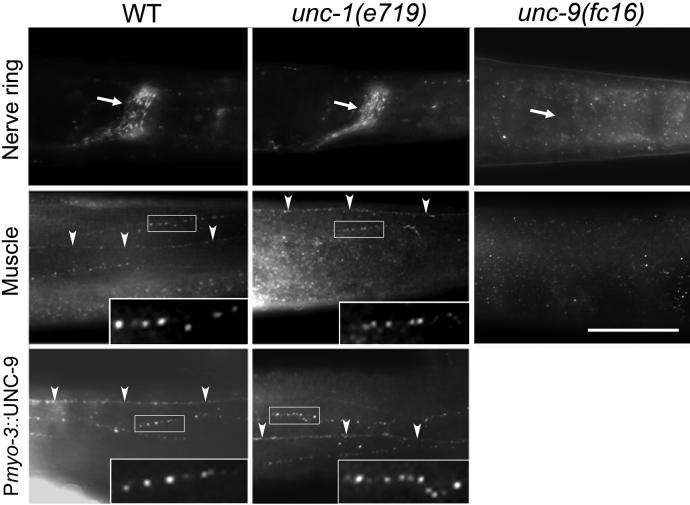
In C. elegans, loss-of-function (lf) mutations of the stomatin-like protein (SLP) UNC-1 and the innexin UNC-9 inhibit locomotion [1, 2] and modulate sensitivity to volatile anesthetics [3, 4]. It was unknown why unc-1(lf) and unc-9(lf) mutants have similar phenotypes. We tested the hypothesis that UNC-1 is a regulator of gap junctions formed by UNC-9. Analyses of junctional currents between body-wall muscle cells showed that electrical coupling was inhibited to a similar degree in unc-1(lf), unc-9(lf), and unc-1(lf);unc-9(lf) double mutants, suggesting that UNC-1 and UNC-9 function together. Expression of Punc-1::DsRED2 and Punc-9::GFP transcriptional fusions suggests that unc-1 and unc-9 are coexpressed in neurons and body-wall muscle cells. Immunohistochemistry showed that UNC-1 and UNC-9 colocalized at intercellular junctions and that unc-1(lf) did not alter UNC-9 expression or subcellular localization. Bimolecular fluorescence complementation (BiFC) assays suggest that UNC-1 and UNC-9 are physically very close at intercellular junctions. Targeted rescue experiments suggest that UNC-9 and UNC-1 function predominantly in neurons to control locomotion. Thus, in addition to the recently reported function of regulating mechanosensitive ion channels [5, 6], SLPs might have a novel function of regulating gap junctions.


Figures

Figure 1
Figure 1
Electrical coupling of body-wall muscle cells was deficient in unc-1 mutants. A: Photograph of a muscle preparation for electrophysiological recording. The two ventral quadrants, with the ventral nerve cord (VNC) and hypodermal ridge (HD ridge) between them, are shown. Each quadrant consists of a monolayer of two rows of muscle cells. Cells in the right quadrant are designated as R1 or R2, whereas those in the left quadrant as L1 or L2. Scale bar = 50 μm. B: Intra-quadrant coupling (between a pair of neighboring R1-R2 or L1-L2 cells) was significantly inhibited in unc-1(fc53) and unc-1(e719). The coupling defect of unc-1(e719) could be rescued by expressing wild-type UNC-1 specifically in body-wall muscle cells under the control of the myosin promoter Pmyo-3. Representative junctional currents and means of the junctional conductance (Gj) are shown. The asterisk indicates a statistically significant difference compared with the wild-type (one-way ANOVA followed by Bonferroni posthoc tests). The number of samples analyzed is indicated by the value above each column.
Figure 2
Figure 2
UNC-1 appeared to be specifically required for the function of UNC-9-based gap junctions in body-wall muscle cells. A: Intra-quadrant coupling (between a pair of neighboring R1-R2 or L1-L2 cells) was indistinguishable between the unc-9(fc16);unc-1(e719) double mutant and either of the single mutant, suggesting that UNC-1 and UNC-9 function in the same pathway. The unc-1(e719) data is the same as that in Figure 1B. B: Wild-type (WT) UNC-9 but not UNC-9::GFP required UNC-1 to rescue electrical coupling of body-wall muscle cells. Inter-quadrant coupling (between a pair of neighboring R1-L1 cells) was nearly absent in unc-9(fc16) or unc-1(e719) mutant. Expression of WT UNC-9 in body-wall muscle cells under the control of Pmyo-3 rescued the coupling defect of unc-9(fc16). In the unc-1(e719) and unc-9(fc16) genetic background, UNC-9::GFP but not WT UNC-9 rescued the coupling defect. The Gj from animals expressing Pmyo-3::UNC-9::GFP might be underestimated because only a small number of progeny expressing the non-integrated transgene survived into adulthood, and these adult animals are conceivably those expressing the transgene at a lower level or in fewer cells. The asterisk indicates a statistically significant difference compared with the WT. The open triangle indicates a statistically significant difference compared with the “unc-9(fc16);unc-1(e719);Pmyo-3::UNC-9” group. One-way ANOVA and Bonferroni posthoc tests were used for the statistical analyses. The number of samples analyzed is indicated by the value above each column. Please note that shown in A and B are intra- and inter-quadrant couplings, respectively, which have different levels of junctional conductance (Gj) and are inhibited to different degrees in unc-9(fc16), as reported previously [7].
Figure 3
Figure 3
UNC-1 and UNC-9 colocalized in body-wall muscle and the nervous system. A: When UNC-1::HA and Myc::UNC-9 were coexpressed specifically in body-wall muscle cells under the control of Pmyo-3, colocalization was observed at body-wall muscle intercellular junctions, muscle arms, and near the ventral and dorsal nerve cords, where muscle arms from the two ventral or dorsal quadrants interdigitate. Immunoreactivity was absent in some muscle cells due to mosaic expression of the non-integrated transgenes. The nerve cord is indicated by arrow heads in the merged picture. Scale bar = 50 μm. B: When Myc::UNC-9 and UNC-1::HA were coexpressed specifically in neurons under the control of Punc-47, colocalization of the two proteins was observed along the nerve cords. Scale bar = 20 μm. C: Coexpression of UNC-1::YFPa (UNC-1 fused to YFP amino terminal) and UNC-9::YFPc (UNC-9 fused to YFP carboxyl terminal) in body-wall muscle cells under the control of Pmyo-3 reconstituted the fluorophore of YFP in vivo. Top panel: Fluorescent puncta were observed at intercellular junctions between muscle cell bodies and between muscle arms along the nerve cord with full-length UNC-1 and UNC-9. A selected region (marked by a rectangular frame) is enlarged and shown above the image (same for the Middle panel). Middle panel: Fluorescent puncta were still observed with deletion of the amino terminal of UNC-1 (amino acid residues 1−167). Lower panel: Deletion of the carboxyl terminal of UNC-1 (amino acid residues 171−289) prevented the BiFC. The bright fluorescent signal in this image was due to autofluorescence of the gut. Scale bar = 50 μm. A note about functionalities of these fusion proteins may be found in the Supplement.
Figure 4
Figure 4
UNC-9 showed normal expression and subcellular localization in unc-1 mutants. Immunostaining was performed in whole-mount worms with an anti-UNC-9 antibody. In wild-type worms, immunoreactive puncta were observed in the nerve ring (indicated by an arrow), along the ventral or dorsal nerve cord (indicated by arrow heads), and between body-wall muscle cell bodies (not labeled). Overexpression of wild-type UNC-9 in body-wall muscle cells under the control of Pmyo-3 caused an enhancement of the immunoreactive puncta. In unc-1(e719) mutant genetic background, UNC-9 expression and subcellular localization were unaltered. In unc-9(fc16) mutant animals, no immunoreactivity was observed, suggesting that the antibody was specific to UNC-9. The nerve cord is marked with arrow heads. Selected regions of UNC-9-immunoreactive puncta (indicated by rectangular frames) are enlarged and shown as insets. Scale bar = 50 μm.

Comment in

References

    1. Brenner S. The genetics of Caenorhabditis elegans. Genetics. 1974;77:71–94. - PMC - PubMed
    1. Park EC, Horvitz HR. Mutations with dominant effects on the behavior and morphology of the nematode Caenorhabditis elegans. Genetics. 1986;113:821–852. - PMC - PubMed
    1. Sedensky MM, Meneely PM. Genetic analysis of halothane sensitivity in Caenorhabditis elegans. Science. 1987;236:952–954. - PubMed
    1. Rajaram S, Sedensky MM, Morgan PG. unc-1: a stomatin homologue controls sensitivity to volatile anesthetics in Caenorhabditis elegans. Proc Natl Acad Sci U S A. 1998;95:8761–8766. - PMC - PubMed
    1. Goodman MB, Ernstrom GG, Chelur DS, O'Hagan R, Yao CA, Chalfie M. MEC-2 regulates C. elegans DEG/ENaC channels needed for mechanosensation. Nature. 2002;415:1039–1042. - PubMed

Publication types

MeSH terms

Substances

Associated data