. 2007 Jul;3(7):e98.
doi: 10.1371/journal.ppat.0030098.

A very late viral protein triggers the lytic release of SV40

Affiliations

A very late viral protein triggers the lytic release of SV40

Robert Daniels et al. PLoS Pathog. 2007 Jul.

Abstract

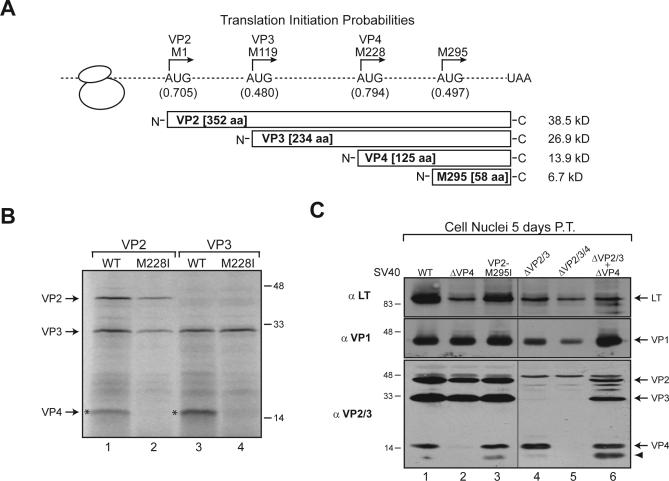
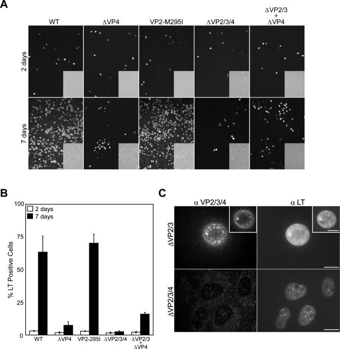
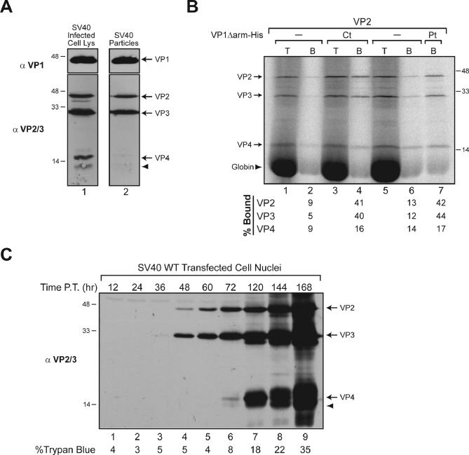
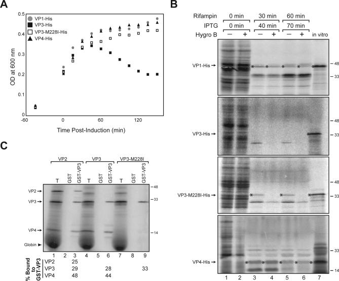
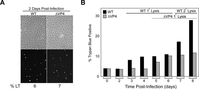
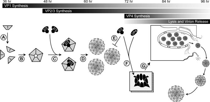
How nonenveloped viruses such as simian virus 40 (SV40) trigger the lytic release of their progeny is poorly understood. Here, we demonstrate that SV40 expresses a novel later protein termed VP4 that triggers the timely lytic release of its progeny. Like VP3, VP4 synthesis initiates from a downstream AUG start codon within the VP2 transcript and localizes to the nucleus. However, VP4 expression occurs approximately 24 h later at a time that coincides with cell lysis, and it is not incorporated into mature virions. Mutation of the VP4 initiation codon from the SV40 genome delayed lysis by 2 d and reduced infectious particle release. Furthermore, the co-expression of VP4 and VP3, but not their individual expression, recapitulated cell lysis in bacteria. Thus, SV40 regulates its life cycle by the later temporal expression of VP4, which results in cell lysis and enables the 50-nm virus to exit the cell. This study also demonstrates how viruses can generate multiple proteins with diverse functions and localizations from a single reading frame.


Conflict of interest statement

Competing interests. The authors have declared that no competing interests exist.

Figures

Figure 1
Figure 1. SV40 Expresses an ∼15-kD Protein Termed VP4
(A) Schematic representation of VP2 displaying the translation initiation probabilities of the in-frame AUG initiation codons as predicted by NetStart1.0 and the respective molecular weights of the resultant polypeptides [12]. (B) VP2 and VP3 mRNAs express VP4 from Met228 in VP2 upon translation in reticulocyte lysate. (C) VP4 is expressed from Met228 in VP2 during SV40 infection. Immunoblot analysis of the cell nuclei fractions isolated from BS-C-1 cells transfected with the designated viral genomes for 72 h and probed with the indicated antibodies. The arrowhead indicates a band with a faster mobility than VP4 that reacts with VP2 and VP3 antisera.
Figure 2
Figure 2. VP4 Is Required for Efficient SV40 Propagation
(A) Representative fluorescence images with the corresponding phase-contrast image (inset) of SV40 propagation in BS-C-1 cells following transfection with WT, ΔVP4, VP2-M295I, and ΔVP2/3/4 genomes and co-transfection with the ΔVP2/3 and ΔVP4 genomes. The cells were fixed at the indicated times post-transfection and stained with LT antisera. (B) Quantifications from two replicate experiments showing the percentage out of ∼3,000 BS-C-1 cells that expressed LT at the indicated times post-transfection with the various SV40 genomes. (C) Fluorescent images of BS-C-1 cells transfected with the ΔVP2/3 or ΔVP2/3/4 genomes for 72 h and stained with antisera for VP2/3/4 (αVP2/3/4) or LT (αLT). Compilations of the z-sections are shown as Max projection images, and insets containing a single z-section from the mid-region of the nucleus are included. The bars represent 10 μm, the antibodies used are noted across the top of the composites, and the transfected genomes are listed to the left.
Figure 3
Figure 3. VP4 Is Expressed at the Onset of Lysis and Is Not Incorporated into SV40 Virions
(A) VP4 is not incorporated into the mature SV40 virions. Immunoblot analysis of SV40-infected cells and isolated virions with the indicated antisera. (B) VP4 does not associate with VP1 pentamers. VP1 pentamers (VP1Δarm-His) purified from bacteria were either present co-translationally (Ct) or during the synthesis of radiolabeled VP2, VP3, and VP4 in reticulocyte lysate or added post-translationally (Pt). The fractions of VP1-bound (B) VP2, VP3, and VP4 were isolated by Ni-NTA-sepharose beads, washed, and resolved by SDS-PAGE next to an equivalent fraction of the total (T) products. The percentage of the bound (B) fraction from two independent experiments is included. (C) Immunoblot analysis of isolated nuclei from BS-C-1 cells at the indicated time post-transfection with the WT SV40 genome demonstrates that VP4 is expressed at the onset of viral-induced host cell lysis. The percentage of ∼2,000 cells staining positive for trypan blue at each time point is listed.
Figure 4
Figure 4. VP4 Expression in Concert with VP3 Induces Bacterial Lysis
(A) E. coli viablility was monitored following the induction of VP1-His, VP3-His, VP3Met228I-His, and VP4-His expression. (B) In the absence of VP4, VP3 renders the E. coli permeable to hygromycin B. Analysis of E. coli permeability to hygromycin B during a 10-min pulse at the indicated time post–IPTG induction in the presence of rifampicin. Radiolabeled in vitro synthesized products of each construct were included as references for the expressed protein. Asterisks denote the overexpressed protein. (C) VP4 oligomerizes with VP3. Radiolabeled VP2, VP3, and VP3-M228I were synthesized in reticulocyte lystate prior to GST-VP3 binding and isolation. The percentage of the total (T) bound to GST-VP3 is indicated.
Figure 5
Figure 5. VP4 Directs the Timely Lytic Release of SV40 Progeny
(A) Representative fluorescence images and the corresponding phase-contrast images displaying WT and ΔVP4 particle infectivity standardization at 2 d. The cells are stained with LT antisera and the percentage of BS-C-1 cells staining for LT is displayed below the images. (B) Removal of VP4 extends the SV40 life cycle 2 d. Trypan blue staining of ∼2,000 BS-C-1 cells at the indicated times post-infection with equivalent WT and ΔVP4 infectious particles.
Figure 6
Figure 6. Proposed Model Displaying the Regulation of the SV40 Lytic Cycle
VP1 is synthesized first, forming pentamers that sequester newly synthesized VP2 and VP3 (steps A–C). Virion assembly (step D) and the stoichiometry of five VP1 per VP2/3 prevent VP2 and VP3 particle incorporation (step E). VP4 is synthesized, it oligomerizes with VP3, and possibly VP2, and targets to the host cell membrane to form a pore (step F), which then initiates the lytic death of the host cell and the release of the progeny (step G).

Similar articles

Cited by

References

    1. Mellman I, Warren G. The road taken: Past and future foundations of membrane traffic. Cell. 2000;100:99–112. - PubMed
    1. Smith AE, Helenius A. How viruses enter animal cells. Science. 2004;304:237–242. - PubMed
    1. Gruenberg J, van der Goot FG. Mechanisms of pathogen entry through the endosomal compartments. Nat Rev Mol Cell Biol. 2006;7:495–504. - PubMed
    1. Kirn D, Martuza RL, Zwiebel J. Replication-selective virotherapy for cancer: Biological principles, risk management and future directions. Nat Med. 2001;7:781–787. - PubMed
    1. van Kuppeveld FJ, Hoenderop JG, Smeets RL, Willems PH, Dijkman HB, et al. Coxsackievirus protein 2B modifies endoplasmic reticulum membrane and plasma membrane permeability and facilitates virus release. EMBO J. 1997;16:3519–3532. - PMC - PubMed

Publication types

MeSH terms