. 2007 Jul;3(7):e103.
doi: 10.1371/journal.ppat.0030103.

Hepatitis C virus p7 protein is crucial for assembly and release of infectious virions

Affiliations

Hepatitis C virus p7 protein is crucial for assembly and release of infectious virions

Eike Steinmann et al. PLoS Pathog. 2007 Jul.

Abstract

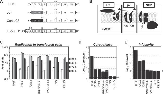
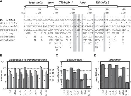
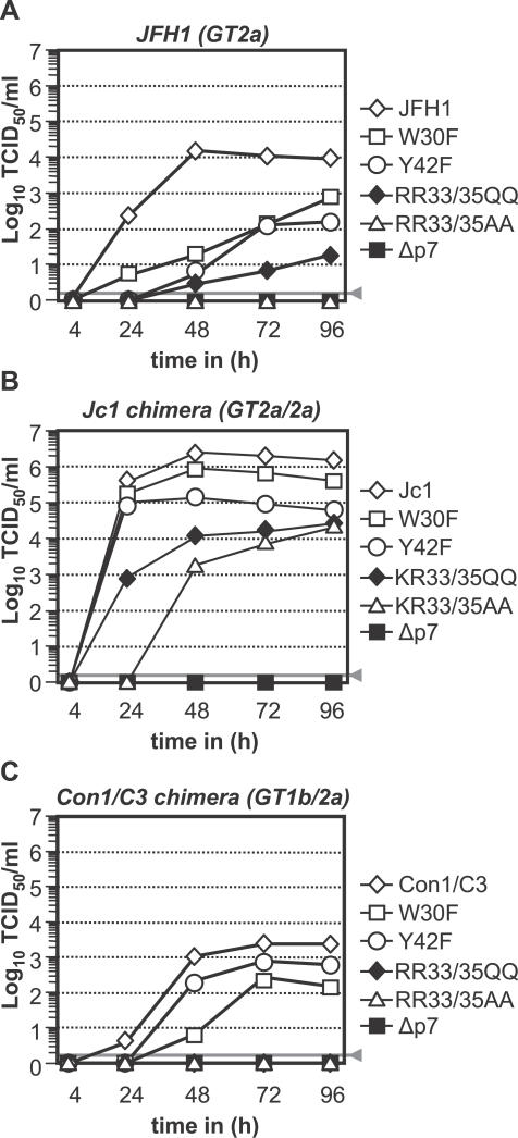
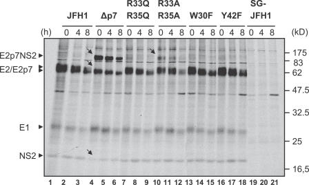
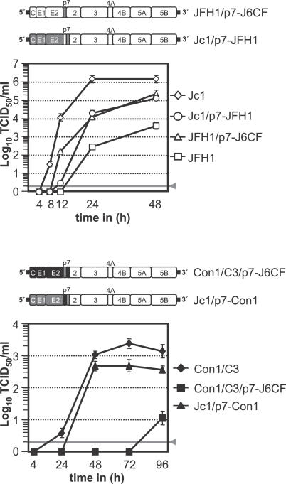
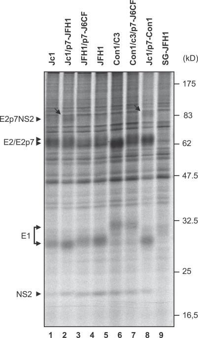
Hepatitis C virus (HCV) infection is associated with chronic liver disease and currently affects about 3% of the world population. Although much has been learned about the function of individual viral proteins, the role of the HCV p7 protein in virus replication is not known. Recent data, however, suggest that it forms ion channels that may be targeted by antiviral compounds. Moreover, this protein was shown to be essential for infectivity in chimpanzee. Employing the novel HCV infection system and using a genetic approach to investigate the function of p7 in the viral replication cycle, we find that this protein is essential for efficient assembly and release of infectious virions across divergent virus strains. We show that p7 promotes virus particle production in a genotype-specific manner most likely due to interactions with other viral factors. Virus entry, on the other hand, is largely independent of p7, as the specific infectivity of released virions with a defect in p7 was not affected. Together, these observations indicate that p7 is primarily involved in the late phase of the HCV replication cycle. Finally, we note that p7 variants from different isolates deviate substantially in their capacity to promote virus production, suggesting that p7 is an important virulence factor that may modulate fitness and in turn virus persistence and pathogenesis.


Conflict of interest statement

Competing interests. The authors have declared that no competing interests exist.

Figures

Figure 1
Figure 1. Replication and Virus Production of Luc-JFH1 Genomes with Mutated p7
(A) Schematic representation of constructs used in this study. JFH1-derived 5′ and 3′ nontranslated regions are drawn as thick black lines and JFH1 proteins are depicted as open boxes. Jc1 and Con1/C3 have been described and comprise chimeric HCV polyproteins consisting of J6CF (gray boxes) or Con1 (black boxes) fused with JFH1 [19]. The luciferase reporter virus genome Luc-JFH1 is depicted at the bottom [21]. (B) Topology model of E2, p7, and NS2 proteins. Signalase cleavage sites are denoted by black arrowheads. The location of conserved di-basic motif of p7 is indicated. C- and N-terminal portion of the E2 TM domain are labeled N and C, respectively, while predicted TM helices 1 and 2 of p7 are labeled 1 and 2. Predicted NS2 TM helix 1 is labeled 1 while the two following predicted helices are labeled 2/3. Note that the crossover site in the virus chimeras is located in the loop following TM helix 1 of NS2 (gray arrow) [19]. (C) Replication of Luc-JFH1 and given mutants in transfected Huh7-Lunet cells determined by luciferase reporter assays. Values given are expressed relative to the reporter activity measured at 4 h which was set to one. Mean values of duplicate measurements and the standard errors are given. (D) Release of core protein into the culture fluid 72 h post-transfection of cell given in (C). The gray lines indicate the cut-off of the ELISA and the background luciferase activity measured in mock-infected cells, respectively; mean values of duplicate measurements and the standard errors are given. (E) Infectivity associated with culture fluids harvested 72 h post-transfection determined by inoculation of Huh-7.5 cells and reporter assays. Mean values of duplicate wells and the standard error of the means are given. The gray line denotes background relative light units measured in mock-infected cells. Data presented in (C–E) were derived from a single experiment representative of five independent repetitions.
Figure 2
Figure 2. Identification of Conserved Amino Acids Essential for p7 Functioning
(A) Amino acid repertoire of the 26 representative p7 sequences of confirmed HCV genotypes/subtypes (listed with accession numbers in Table 1 in [1]). Helical turn and loop regions deduced from the nuclear magnetic resonance structure analysis of a p7 GT 1b variant (F. Penin, unpublished data) are depicted at the top, and the primary sequence of JFH1-derived p7 (GT 2a) in combination with a ruler indicating amino acid numbers with respect to p7 and the complete JFH1 genome are given below. The degree of conservation can be inferred by the extent of variability (with the observed amino acids listed in decreasing order of frequency from top to bottom) and the similarity index according to ClustalW convention (asterisk, invariant; colon, highly similar; dot, similar) [46]. Residues mutated in this study are shaded in gray. (B) Replication of Luc-JFH1 and given mutants in transfected Huh7-Lunet cells determined as described above. (C) Release of core protein and (D) infectivity into the culture fluid 72 h post-transfection of cell given in (B). The gray lines indicate the cut-off of the ELISA and the background luciferase activity measured in mock-infected cells, respectively. A representative experiment of at least four independent repetitions is given.
Figure 3
Figure 3. Accumulation of Infectivity upon Transfection of JFH1 or Chimeras Carrying Varying p7 Mutations
Huh-7.5 cells were transfected with (A) JFH1, (B) Jc1, or (C) Con1/C3 wild-type constructs or given p7 mutants in the context of the respective virus. Cell-free supernatants were harvested and titrated by using a limiting dilution assay. The gray bars represent the detection limit of the limiting dilution assay.
Figure 4
Figure 4. Processing of the E2-p7-NS2 Region of JFH1 and Various p7 Mutants
Huh-7.5 cells were transfected with indicated constructs and after 24 h pulse-labeled with [35S] methionine/cysteine-containing medium, lysed immediately, or chased for 4 or 8 h. Cells transfected with a subgenomic JFH1 replicon encoding HCV proteins NS3–NS5B only (SG-JFH1) served as negative control. HCV proteins were immunoprecipitated using an antibody monospecific for E2 and are identified by arrows on the left. The positions of the molecular weight standard are given on the right. Arrowheads to the left point to the respective precursors and mature proteins detected. Arrows within the figure point out obvious alterations in protein processing.
Figure 5
Figure 5. Specific Infectivity and Efficiency of Virus Assembly and Release of Jc1 with Mutated p7
(A) Huh7-Lunet cells were transfected with indicated genomes, and 48 h post-transfection culture fluid was harvested for quantification of core protein by ELISA (left panel) and infectivity by TCID50 assay (middle). The specific infectivity of the given viruses was determined by dilution of Jc1 and ΔHVR1 to the same quantity of core protein as KR33/35QQ (444 pg) and titration by using the limiting dilution assay. (B) Cells were transfected as in (A) and 48 h post-transfection supernatants were collected. In parallel, virus-producing cells were washed and lysed by repetitive cycles of freeze and thaw. Extracellular (white bars) and intracellular infectivity (gray bars) were determined by limiting dilution assay and are depicted in the middle. The panel on the left shows the total infectivity per plate, whereas the percentage of cell-associated and released infectivity expressed relative to the total amount of infectivity is given in the right. The gray lines denote the detection limits of the ELISA and the limiting dilution assay, respectively. Mean values of five independent experiments and the standard deviation of the means are presented.
Figure 6
Figure 6. Accumulation of Infectivity upon Transfection of GT 2a and GT 1b Viruses with J6CF-Derived p7 Proteins
The color coding is as in Figure 1 with open boxes representing JFH1, gray boxes J6CF, and black boxes Con1 proteins. Accumulation of infectivity upon transfection is given below. The gray line denotes the detection limit of the limiting dilution assay. A representative experiment of at least two repetitions is depicted.
Figure 7
Figure 7. Processing of the E2-p7-NS2 Region of JFH1, Jc1, Con1/C3, and Various Chimeras
Huh-7.5 cells were transfected with indicated constructs. 24 h post-transfection, cells were labeled with [35S] methionine/cysteine-containing medium overnight and then lysed immediately. HCV proteins were immunoprecipitated using an antibody monospecific for E2. The positions of the molecular weight standard are given on the right. Arrowheads to the left point to the respective precursors and mature HCV proteins detected. Arrows within the figure point out alterations in protein processing. Note that Con1-derived E1 displays a lower electrophoretic mobility than E1 from JFH1 and J6CF.

References

    1. Simmonds P, Bukh J, Combet C, Deleage G, Enomoto N, et al. Consensus proposals for a unified system of nomenclature of hepatitis C virus genotypes. Hepatology. 2005;42:962–973. - PubMed
    1. Hoofnagle JH. Course and outcome of hepatitis C. Hepatology. 2002;36:S21–S29. - PubMed
    1. Manns MP, Wedemeyer H, Cornberg M. Treating viral hepatitis C: Efficacy, side effects, and complications. Gut. 2006;55:1350–1359. - PMC - PubMed
    1. Bartenschlager R, Frese M, Pietschmann T. Novel insights into hepatitis C virus replication and persistence. Adv Virus Res. 2004;63:71–180. - PubMed
    1. Penin F, Dubuisson J, Rey FA, Moradpour D, Pawlotsky JM. Structural biology of hepatitis C virus. Hepatology. 2004;39:5–19. - PubMed

Publication types

Associated data