. 2007 Jul;3(7):e110.
doi: 10.1371/journal.ppat.0030110.

Mycobacterium tuberculosis nuoG is a virulence gene that inhibits apoptosis of infected host cells

Affiliations

Mycobacterium tuberculosis nuoG is a virulence gene that inhibits apoptosis of infected host cells

Kamalakannan Velmurugan et al. PLoS Pathog. 2007 Jul.

Abstract

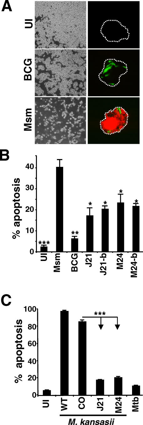
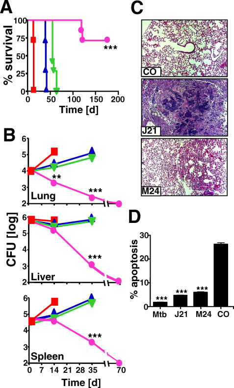
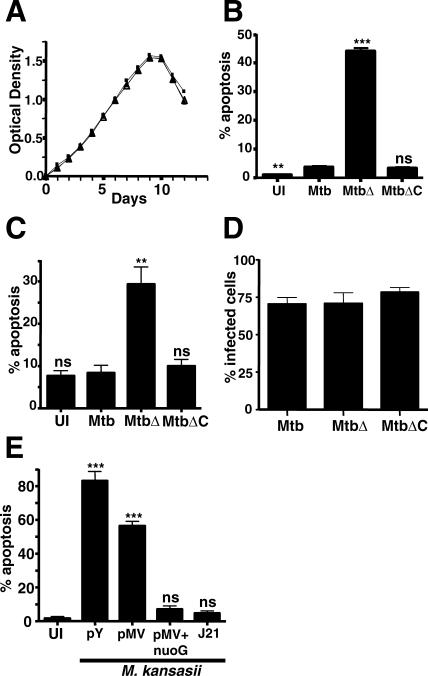
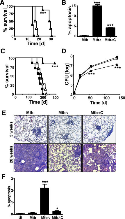
The survival and persistence of Mycobacterium tuberculosis depends on its capacity to manipulate multiple host defense pathways, including the ability to actively inhibit the death by apoptosis of infected host cells. The genetic basis for this anti-apoptotic activity and its implication for mycobacterial virulence have not been demonstrated or elucidated. Using a novel gain-of-function genetic screen, we demonstrated that inhibition of infection-induced apoptosis of macrophages is controlled by multiple genetic loci in M. tuberculosis. Characterization of one of these loci in detail revealed that the anti-apoptosis activity was attributable to the type I NADH-dehydrogenase of M. tuberculosis, and was mainly due to the subunit of this multicomponent complex encoded by the nuoG gene. Expression of M. tuberculosis nuoG in nonpathogenic mycobacteria endowed them with the ability to inhibit apoptosis of infected human or mouse macrophages, and increased their virulence in a SCID mouse model. Conversely, deletion of nuoG in M. tuberculosis ablated its ability to inhibit macrophage apoptosis and significantly reduced its virulence in mice. These results identify a key component of the genetic basis for an important virulence trait of M. tuberculosis and support a direct causal relationship between virulence of pathogenic mycobacteria and their ability to inhibit macrophage apoptosis.


Conflict of interest statement

Competing interests. The authors have declared that no competing interests exist.

Figures

Figure 1
Figure 1. Identification of Regions in the Mtb Genome Mediating Inhibition of THP-1 Cell Apoptosis
(A) M. smegmatis (Msm) induced more cell death in infected THP-1 cells than BCG or uninfected cells (UI) as observed by bright field microscopy (left panels) and fluorescence microscopy of TUNEL staining (right panels; red fluorescence is TUNEL staining, and green fluorescence is GFP-labeled bacteria). (B and C) M. smegmatis was transfected with an episomal cosmid library of Mtb genomic DNA, and individual clones were screened for their capacity to inhibit apoptosis. The cosmid DNA of two selected clones (J21 and M24) was purified and used to re-transfect M. smegmatis, resulting in clones J21-b and M24-b. These clones, along with the original transformants (M24 and J21), uninfected (UI) cells, M. smegmatis (Msm), and BCG were tested for induction of apoptosis by infection of THP-1 cells followed by TUNEL staining at 16 h after infection and flow cytometry (C). The J21 and M24 cosmids and empty vector cosmid (CO) were transfected into M. kansasii, and the induction of apoptosis by the bacteria was compared to uninfected and Mtb-infected THP-1 cells using a TUNEL assay as in (B), except that cells were harvested after 5 d of infection. Results in (B and C) are averages of three independent experiments and error bars represent ± standard deviation (SD). Statistical significance relative to levels of apoptosis induced by wild-type M. smegmatis in (B) or M. kansasii in (C) is indicated as follows: *, 0.01 < p < 0.05; **, 0.001 < p < 0.01; ***, p < 0.001 (ANOVA with Tukey post-test).
Figure 2
Figure 2. Correlation between Apoptosis Inhibition and Mycobacterial Virulence
(A and B) SCID mice were infected intravenously with 106 bacteria and survival ([A], n = 7 per group), or the bacterial load in lung, liver, and spleen ([B], n = 3 per time point), were determined. Shown are results with Mtb (squares), Mkan-J21 (triangle), Mkan-M24 (inverted triangle), and Mkan-CO (circle). (C) Histopathology (hematoxylin and eosin staining) of lungs from SCID mice infected 5 wk earlier with Mkan-CO, Mkan-J21, or Mkan-M24 as indicated. (D) Levels of apoptosis in vivo after 2 wk of infection were determined by quantification of TUNEL-positive cells per total cell nuclei in lung tissue sections. Means ±SD for blinded analysis of about 2,000 total cells per tissue section (three sections per lung) per mouse (n = 3) are shown. The results shown are representative of three independent experiments. Error bars representing ±SD are shown in (D) and are smaller than symbols in (B). Asterisks indicate p-values as in Figure 1.
Figure 3
Figure 3. Importance of Mtb nuoG for Inhibition of Macrophage Apoptosis
(A) The in vitro growth of wild-type (squares), mutant (full triangles), and complemented bacteria (empty triangles) was analyzed at the indicated time points in triplicate cultures (error bars showing ±SD are smaller than symbols). (B and C) Induction of apoptosis by infection of differentiated human THP-1 cells (B) or cultured primary mouse macrophages (C) with wild-type (Mtb), the ΔnuoG deletion mutant (MtbΔ), or complemented mutant (MtbΔC). Cells were harvested either at day 3 (B) or day 1 (C) after infection, stained by TUNEL assay, and analyzed using flow cytometry. (D) The percentage of infected macrophages in (C) was determined by acid-fast staining. (E) M. kansasii was transfected with empty cosmid (pY), empty plasmid (pMV), cosmid J21 (J21), and Mtb nuoG behind the constitutive promoter of pMV261 (pMV+nuoG), and induction of apoptosis in THP-1 cells was determined by TUNEL staining and flow cytometry. All results shown are means ±SD of triplicate cultures and are representative of three independent experiments. Asterisks indicate statistical significance as in Figure 1. ns, not significant.
Figure 4
Figure 4. Identification of nuoG as a Mycobacterial Virulence Determinant
(A) Survival of SCID mice after intravenous infection with 106 wild-type Mtb (squares), ΔnuoG mutant (filled triangles), or complemented mutant bacilli (open triangles). n = 7 mice per group. (B) Apoptotic cells in lung tissues of SCID mice after 14 d of infection were quantified using TUNEL peroxidase staining by microscopy as explained in Figure 2. (C) Survival of immunocompetent BALB/c mice infected with bacterial strains as in (A); n = 7 mice per group. (D) The bacterial burden in the lungs of infected BALB/c mice was followed (n = 3 per time point; symbols indicate bacterial strains as in [A and C]). (E) Lung histopathology (hematoxylin and eosin staining) at 3 and 20 wk for BALB/c mice infected as in (C) with wild-type Mtb, ΔnuoG mutant (MtbΔ), or complemented mutant Mtb (MtbΔC). (F) The percentages of apoptotic cells in lung sections were determined by TUNEL peroxidase assay and quantified in blinded fashion as in Figure 2. All results shown are representative of two independent experiments. Statistically significant differences compared to wild-type Mtb are indicated by asterisks as in the Figure 1 legend.

References

    1. Dye C, Watt CJ, Bleed DM, Hosseini SM, Raviglione MC. Evolution of tuberculosis control and prospects for reducing tuberculosis incidence, prevalence, and deaths globally. JAMA. 2005;293:2767–2775. - PubMed
    1. Manabe YC, Bishai WR. Latent Mycobacterium tuberculosis—Persistence, patience, and winning by waiting. Nat Med. 2000;6:1327–1329. - PubMed
    1. Honer zu Bentrup K, Russell DG. Mycobacterial persistence: Adaptation to a changing environment. Trends Microbiol. 2001;9:597–605. - PubMed
    1. Hingley-Wilson SM, Sambandamurthy VK, Jacobs WR., Jr Survival perspectives from the world's most successful pathogen, Mycobacterium tuberculosis . Nat Immunol. 2003;4:949–955. - PubMed
    1. Smith CV, Sharma V, Sacchettini JC. TB drug discovery: Addressing issues of persistence and resistance. Tuberculosis (Edinb) 2004;84:45–55. - PubMed

Publication types

MeSH terms

Associated data