Comparative Study
. 2007 Jul 20:5:31.
doi: 10.1186/1477-7827-5-31.

Prostaglandin F2-alpha receptor (FPr) expression on porcine corpus luteum microvascular endothelial cells (pCL-MVECs)

Affiliations
Comparative Study

Prostaglandin F2-alpha receptor (FPr) expression on porcine corpus luteum microvascular endothelial cells (pCL-MVECs)

Augusta Zannoni et al. Reprod Biol Endocrinol. .

Abstract

Background: The corpus luteum (CL) is a transient endocrine gland and prostaglandin F2-alpha is considered to be the principal luteolysin in pigs. In this species, the in vivo administration of prostaglandin F2-alpha induces apoptosis in large vessels as early as 6 hours after administration. The presence of the prostaglandin F2-alpha receptor (FPr) on the microvascular endothelial cells (pCL-MVECs) of the porcine corpus luteum has not yet been defined. The aim of the study was to assess FPr expression in pCL-MVECs in the early and mid-luteal phases (EL-p, ML-p), and during pregnancy (P-p). Moreover, the effectiveness of prostaglandin F2-alpha treatment in inducing pCL-MVEC apoptosis was tested.

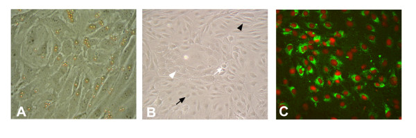
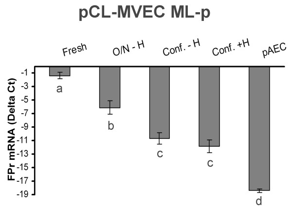
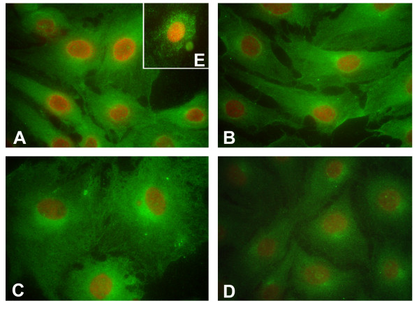
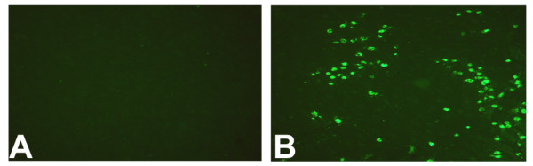
Methods: Porcine CLs were collected in the EL and ML phases and during P-p. All CLs from each animal were minced together and the homogenates underwent enzymatic digestion. The pCL-MVECs were then positively selected by an immunomagnetic separation protocol using Dynabeads coated with anti-CD31 monoclonal antibody and seeded in flasks in the presence of EGM 2-MV (Microvascular Endothelial Cell Medium-2). After 4 days of culture, the cells underwent additional immunomagnetic selection and were seeded in flasks until the confluent stage.PCR Real time, western blot and immunodetection assays were utilized to assess the presence of FPr on pCL-MVEC primary cultures. Furthermore, the influence of culture time (freshly isolated, cultured overnight and at confluence) and hormonal treatment (P4 and E2) on FPr expression in pCL-MVECs was also investigated. Apoptosis was detected by TUNEL assay of pCL-MVECs exposed to prostaglandin F2-alpha.

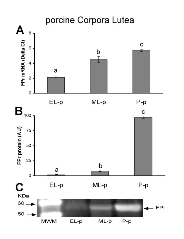
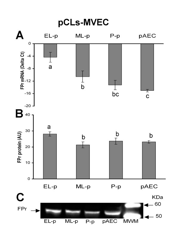
Results: We obtained primary cultures of pCL-MVECs from all animals. FPr mRNA and protein levels showed the highest value (ANOVA) in CL-MVECs derived from the early-luteal phase. Moreover, freshly isolated MVECs showed a higher FPr mRNA value than those cultured overnight and confluent cells (ANOVA). prostaglandin F2-alpha treatment failed to induce an apoptotic response in all the pCL-MVEC cultures.

Conclusion: Our data showing the presence of FPr on MVECs and the inability of prostaglandin F2-alpha to evoke an in vitro apoptotic response suggest that other molecules or mechanisms must be considered in order to explain the in vivo direct pro-apoptotic effect of prostaglandin F2-alpha at the endothelial level.


Figures

Figure 1
Figure 1
Representative images of mid-uteal-phase porcine corpus luteum-microvascular endothelial cell (ML-p pCL-MVEC) cultures. A) pCL-MVECs after 2 days of culture with beads still attached (phase contrast microscope ×200). B) the pCL-MVEC monolayer at confluence (day 7) (phase contrast microscope ×100). Different endothelial cell types are shown: spindle-shaped cells (black arrowhead), cobble-stone cells (white arrow), polygonal-opaque cells (black arrow), phase-dense cells (white arrowhead). C) Immunostaining for FVIII antigen (epifluorescence microscope ×100).
Figure 2
Figure 2
Presence of FPr in porcine corpora lutea (pCLs) at the early-luteal phase (EL-p), the mid-luteal phase (ML-p) and during pregnancy (P-p). A) Relative FPr mRNA expression. The results are presented as Delta Ct (HPRT Ct - FPr Ct). B) FPr protein content. The results are presented as AU (arbitrary units). Data represent mean ± SD. Different letters indicate statistically significant differences (P < 0.05). C) Representative Western blotting of FPr.
Figure 3
Figure 3
Presence of FPr in porcine corpora lutea-derived microvascular endothelial cells (pCL-MVECs) and porcine Aortic Endothelial Cells (pAECs). A) Relative FPr mRNA expression. The results are presented as Delta Ct (HPRT Ct - FPr Ct). B) FPr protein content. The results are presented as AU (arbitrary units). Data represent mean ± SD. Different letters indicate statistically significant differences (P < 0.05). C) Representative Western blotting of FPr.
Figure 4
Figure 4
FPr mRNA expression in porcine corpora lutea-microvascular endothelial cells (pCL-MVECs) freshly isolated (Fresh) and cultured for different times (overnight-O/N; confluence-Conf.) and in porcine Aortic Endothelial Cells (pAECs). The confluent stage of the MVECs was reached in the presence (+) or absence (-) of hormonal treatment (H = P4 [10-6M] + E2 [10-8 M]). The results are presented as Delta Ct (HPRT Ct - FPr Ct). The data represent mean ± SD. Different letters indicate statistically significant differences (P < 0.05).
Figure 5
Figure 5
Representative FPr immunofluorescent staining on porcine corpus luteum-microvascular endothelial cells (pCL-MVECs) isolated from the early luteal phase (EL-p) (A), the mid-luteal phase (ML-p) (B) and during pregnancy (P-p) (C) (×400). D) porcine Aortic Endothelial Cell (pAEC) culture (x400). Representative immunostaining of the FPr on non-fixed EL-p pCL-MVECs is shown (E).
Figure 6
Figure 6
Effect of PGFtreatment on apoptosis induction in porcine corpus luteum-microvascular endothelial cells (pCL-MVECs). A) Representative images of a TUNEL assay in PGF-treated mid-luteal phase (ML-p) pCL-MVECs. B) Positive TUNEL control (LPS-treated pAECs) (×100).

Similar articles

Cited by

References

    1. Niswender GD, Juengel JL, Silva PJ, Rollyson MK, McIntush EW. Mechanisms controlling the function and life span of the corpus luteum. Physiol Rev. 2000;80:1–29. - PubMed
    1. Balapure AK, Caicedo IC, Kawada K, Watt DS, Rexroad CE, Jr, Fitz TA. Multiple classes of prostaglandin F2 alpha binding sites in subpopulations of ovine luteal cells. Biol Reprod. 1989;41:385–392. doi: 10.1095/biolreprod41.3.385. - DOI - PubMed
    1. Gadsby JE, Balapure AK, Britt JH, Fitz TA. Prostaglandin F2 alpha receptors on enzyme-dissociated pig luteal cells throughout the estrous cycle. Endocrinology. 1990;126:787–795. - PubMed
    1. Sakamoto K, Kamimura M, Kurozumi S, Ito S. Prostaglandin F2 alpha receptor. J Lipid Media Cell Signal. 1995;12:405–411. doi: 10.1016/0929-7855(95)00026-M. - DOI - PubMed
    1. Juengel JL, Wiltbank MC, Meberg BM, Niswender GD. Regulation of steady-state concentrations of messenger ribonucleic acid encoding prostaglandin F2 alpha receptor in ovine corpus luteum. Biol Reprod. 1996;54:1096–1102. doi: 10.1095/biolreprod54.5.1096. - DOI - PubMed

Publication types

LinkOut - more resources