. 2007;40(16):3660-71.
doi: 10.1016/j.jbiomech.2007.06.006. Epub 2007 Jul 19.

Contributions of muscles to terminal-swing knee motions vary with walking speed

Affiliations

Contributions of muscles to terminal-swing knee motions vary with walking speed

Allison S Arnold et al. J Biomech. 2007.

Abstract

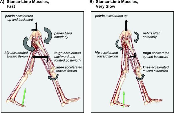
Many children with cerebral palsy walk with diminished knee extension during terminal swing, at speeds much slower than unimpaired children. Treatment of these gait abnormalities is challenging because the factors that extend the knee during normal walking, over a range of speeds, are not well understood. This study analyzed a series of three-dimensional, muscle-driven dynamic simulations to determine whether the relative contributions of individual muscles and other factors to angular motions of the swing-limb knee vary with walking speed. Simulations were developed that reproduced the measured gait dynamics of seven unimpaired children walking at self-selected, fast, slow, and very slow speeds (7 subjects x 4 speeds=28 simulations). In mid-swing, muscles on the stance limb made the largest net contribution to extension of the swing-limb knee at all speeds examined. The stance-limb hip abductors, in particular, accelerated the pelvis upward, inducing reaction forces at the swing-limb hip that powerfully extended the knee. Velocity-related forces (i.e., Coriolis and centrifugal forces) also contributed to knee extension in mid-swing, though these contributions were diminished at slower speeds. In terminal swing, the hip flexors and other muscles on the swing-limb decelerated knee extension at the subjects' self-selected, slow, and very slow speeds, but had only a minimal net effect on knee motions at the fastest speeds. Muscles on the stance limb helped brake knee extension at the subjects' fastest speeds, but induced a net knee extension acceleration at the slowest speeds. These data--which show that the contributions of muscular and velocity-related forces to terminal-swing knee motions vary systematically with walking speed--emphasize the need for speed-matched control subjects when attempting to determine the causes of a patient's abnormal gait.


Figures

Figure 1
Figure 1
Fast, self-selected, slow, and very slow walking speeds of the subjects in this study. Simulations were created that reproduced the gait dynamics of each subject at each speed.
Figure 2
Figure 2
Muscle-driven simulations of swing phase that reproduce the gait dynamics of a 13-year-old subject, Subject 7, walking at fast (1.4 m/s), self-selected (1.0 m/s), slow (0.7 m/s), and very slow (0.5 m/s) speeds. To create these simulations, a musculoskeletal model with 21 degrees of freedom and 92 muscles was scaled to the subject's mass (41.6 kg) and height (1.6 m). Each simulation is shown at the instants just prior to toe-off (left), just prior to initial contact (right), and at peak swing-phase knee flexion (center).
Figure 3
Figure 3
Activation patterns for 10 of the 92 muscle-tendon actuators on the swing limb (left) and stance limb (right) used to drive the simulations of an 11-year-old subject, Subject 4, at fast, self-selected, slow, and very slow walking speeds (darker lines correspond to faster speeds). Note that our model has the isometric force-generating capacity of an adult, while the subject has the mass (31.7 kg) and height (1.4 m) of a child. The magnitudes of the muscle activations, therefore, reflect the relatively small activations (and forces) needed to track the subject's gait dynamics. Corresponding EMG on/off times published by Perry (1992) are overlaid for comparison (solid bars; the thinner bars indicate inconsistencies in EMG timing as documented by Perry), and are scaled to the subject's measured stance and swing phases at the self-selected speed.
Figure 4
Figure 4
Knee flexion angle (A) and angular acceleration (B) during the swing phase as determined experimentally for a 14-year-old subject, Subject 5, walking at fast (1.5 m/s), self-selected (1.1 m/s), slow (0.7 m/s), and very slow (0.5 m/s) speeds. Prior to toe-off, the knee is rapidly accelerated toward flexion. Near toe-off, the knee stops accelerating toward flexion and starts accelerating toward extension due to the actions of muscles and velocity-related forces. In late swing, the knee stops accelerating toward extension and starts accelerating toward flexion due to the actions of muscles. The extension phase is defined as the interval during which the knee is accelerated toward extension; the braking phase is defined as the interval during which the knee is accelerated toward flexion.
Figure 5
Figure 5
Angular acceleration of the swing-limb knee induced by gravity, velocity-related forces, and muscles, averaged over the extension phase (A) and the braking phase (B), at fast, self-selected, slow, and very slow walking speeds. Each bar represents the mean + 1 SD for the seven subjects in this study.
Figure 6
Figure 6
Angular acceleration of the swing-limb knee induced by swing-limb muscles and stance-limb muscles, averaged over the extension phase (A) and the braking phase (B), at fast, self-selected, slow, and very slow walking speeds. Each bar represents the mean + 1 SD for the seven subjects in this study. The net knee accelerations induced by all muscles (swing-limb muscles, stance-limb muscles, and back muscles) are shown for comparison.
Figure 7
Figure 7
Angular acceleration of the swing-limb knee induced by individual muscles or groups of muscles on the swing limb (light grey bars) and on the stance limb (dark grey bars), averaged over the extension phase at fast (A) and very slow speeds (B). DF, the ankle dorsiflexors, includes tibialis anterior, extensor digitorum longus, extensor hallucis longus, and peroneus tertius. HipFlx, the hip flexors, includes iliacus, psoas, tensor fasciae latae, and sartorius. BFSH is the biceps femoris short head. RF is the rectus femoris. VAS includes vastus medialis, vastus intermedius, and vastus lateralis. UPF, the uniarticular ankle plantarflexors, includes soleus, tibialis posterior, flexor digitorum longus, flexor hallucis longus, peroneus longus, and peroneus brevis. HipExt, the stance-limb hip extensors, includes gluteus maximus, hamstrings, and adductor magnus. HipAb, the stance-limb hip abductors, includes gluteus medius and gluteus minimus. Other includes all other muscles of the corresponding limb in the model.
Figure 8
Figure 8
Angular acceleration of the swing-limb knee during the braking phase induced by swing-limb muscles (A) and stance-limb muscles (B), expressed as a percentage of the total knee acceleration and plotted vs. walking speed. Data for each of the seven subjects are shown. Swing-limb muscles exert a greater net influence on the knee motions at slow speeds, while stance-limb muscles exert a greater net influence on the knee motions at fast speeds.
Figure 9
Figure 9
Angular acceleration of the swing-limb knee induced by individual muscles or groups of muscles on the swing limb (light grey bars) and on the stance limb (dark grey bars), averaged over the braking phase at fast (A) and very slow speeds (B). DF, the ankle dorsiflexors, includes tibialis anterior, extensor digitorum longus, extensor hallucis longus, and peroneus tertius. HipFlx, the hip flexors, includes iliacus, psoas, tensor fasciae latae, and sartorius. HAMS, the hamstrings, includes semimembranosus, semitendinosus, and biceps femoris long head. VAS includes vastus medialis, vastus intermedius, and vastus lateralis. UPF, the uniarticular ankle plantarflexors, includes soleus, tibialis posterior, flexor digitorum longus, flexor hallucis longus, peroneus longus, and peroneus brevis. HipExt, the swing-limb hip extensors, includes gluteus maximus and adductor magnus. HipAb, the stance-limb hip abductors, includes gluteus medius and gluteus minimus. Other includes all other muscles of the corresponding limb in the model.
Figure 10
Figure 10
Motions of the pelvis and swing limb induced by all stance-limb muscles during the braking phase at fast (A) and very slow speeds (B). Straight arrows represent translational accelerations, and curved arrows represent angular accelerations. All arrows are scaled proportional to their magnitudes. Accelerations of the thigh are calculated relative to the pelvis. Stance-limb muscles accelerated the model's center of mass (not shown) upward and forward during the braking phase, consistent with previous studies (Liu et al., 2006).
Figure 11
Figure 11
Motions of the pelvis and swing limb induced by all swing-limb muscles during the braking phase at fast (A) and very slow speeds (B). Straight arrows represent translational accelerations, and curved arrows represent angular accelerations. All arrows are scaled proportional to their magnitudes. Accelerations of the thigh are calculated relative to the pelvis, and accelerations of the shank are calculated relative to the thigh.

References

    1. Anderson FC, Goldberg SR, Pandy MG, Delp SL. Contributions of muscle forces and toe-off kinematics to peak knee flexion during the swing phase of normal gait: an induced position analysis. Journal of Biomechanics. 2004;37:731–737. - PubMed
    1. Andersson EA, Nilsson J, Thorstensson A. Intramuscular EMG from the hip flexor muscles during human locomotion. Acta Physiologica Scandinavica. 1997;161:361–370. - PubMed
    1. Arnold AS, Anderson FC, Pandy MG, Delp SL. Muscular contributions to hip and knee extension during the single limb stance phase of normal gait: a framework for investigating the causes of crouch gait. Journal of Biomechanics. 2005;38:2181–2189. - PubMed
    1. Arnold AS, Thelen DG, Schwartz MH, Anderson FC, Delp SL. Muscular coordination of knee motion during the terminal swing phase of gait. Journal of Biomechanics. in press. - PMC - PubMed
    1. Chen G. Induced acceleration contributions to locomotion dynamics are not physically well defined. Gait and Posture. 2006;23:37–44. - PubMed

Publication types