

A gastrointestinal stromal tumor of the duodenum masquerading as a pancreatic head tumor
- PMID: 17659684
- PMCID: PMC4172725
- DOI: 10.3748/wjg.v13.i24.3396
A gastrointestinal stromal tumor of the duodenum masquerading as a pancreatic head tumor
Abstract
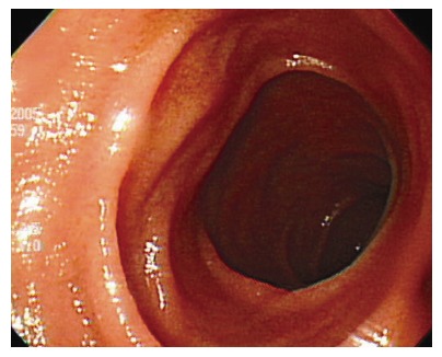
Gastrointestinal stromal tumor (GIST) represents the most common kind of mesenchymal tumor that arises from the alimentary tract. GIST is currently defined as a gastrointestinal tract mesenchymal tumor showing CD117 (c-kit protein) positivity at immunohistochemistry. Throughout the whole length of the gastrointestinal tract, GIST arises most commonly from the stomach followed by the small intestine, the colorectum, and the esophagus. Only 3%-5% of GISTs occur in the duodenum, and especially, if GIST arises from the C loop of the duodenum, it can be difficult to differentiate from the pancreas head mass because of its anatomical proximity. Here, we report a case of duodenal GIST, which was assessed as a pancreatic head tumor preoperatively.
Figures


References
-
- Miettinen M, Lasota J. Gastrointestinal stromal tumors--definition, clinical, histological, immunohistochemical, and molecular genetic features and differential diagnosis. Virchows Arch. 2001;438:1–12. - PubMed
-
- Fletcher CD, Berman JJ, Corless C, Gorstein F, Lasota J, Longley BJ, Miettinen M, O'Leary TJ, Remotti H, Rubin BP, et al. Diagnosis of gastrointestinal stromal tumors: A consensus approach. Hum Pathol. 2002;33:459–465. - PubMed
-
- Joensuu H, Fletcher C, Dimitrijevic S, Silberman S, Roberts P, Demetri G. Management of malignant gastrointestinal stromal tumours. Lancet Oncol. 2002;3:655–664. - PubMed
-
- Miettinen M, Kopczynski J, Makhlouf HR, Sarlomo-Rikala M, Gyorffy H, Burke A, Sobin LH, Lasota J. Gastrointestinal stromal tumors, intramural leiomyomas, and leiomyosarcomas in the duodenum: a clinicopathologic, immunohistochemical, and molecular genetic study of 167 cases. Am J Surg Pathol. 2003;27:625–641. - PubMed
Publication types
MeSH terms
LinkOut - more resources
Full Text Sources
Medical
