Relationship of hepatitis B virus infection of placental barrier and hepatitis B virus intra-uterine transmission mechanism
- PMID: 17659715
- PMCID: PMC4146804
- DOI: 10.3748/wjg.v13.i26.3625
Relationship of hepatitis B virus infection of placental barrier and hepatitis B virus intra-uterine transmission mechanism
Abstract
Aim: To explore the mechanism of intra-uterine transmission, the HBV infection status of placental tissue and in vitro cultured placental trophoblastic cells was tested through in vivo and in vitro experiments.
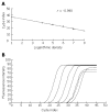
Methods: A variety of methods, such as ELISA, RT-PCR, IHC staining and immunofluorescent staining were employed to test the HBV marker positive pregnant women's placenta and in vitro cultured placental trophoblastic cells.
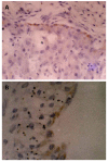
Results: The HBV DNA levels in pregnant women's serum and fetal cord blood were correlated. For those cord blood samples positive for HBV DNA, their maternal blood levels of HBV DNA were at a high level. The HBsAg IHC staining positive cells could be seen in the placental tissues and the presence of HBV DNA detected. After co-incubating the trophoblastic cells and HBV DNA positive serum in vitro, the expressions of both HBsAg and HBV DNA could be detected.
Conclusion: The mechanism of HBV intra-uterine infection may be due to that HBV breaches the placental barrier and infects the fetus.
Figures


References
-
- Bai H, Zhao GZ. Progresses in the study on the mechanism of HBV transmission from mother to infant. Guowai Yixue Liuxingbingxue Chuanranbingxue Fence. 2005;32:99–102.
-
- Michielsen PP, Van Damme P. Viral hepatitis and pregnancy. Acta Gastroenterol Belg. 1999;62:21–29. - PubMed
-
- van der Sande MA, Waight P, Mendy M, Rayco-Solon P, Hutt P, Fulford T, Doherty C, McConkey SJ, Jeffries D, Hall AJ, et al. Long-term protection against carriage of hepatitis B virus after infant vaccination. J Infect Dis. 2006;193:1528–1535. - PubMed
-
- Kripke C. Hepatitis B vaccine for infants of HBsAg-positive mothers. Am Fam Physician. 2007;75:49–50. - PubMed
-
- Xiao XM, Li AZ, Chen X, Zhu YK, Miao J. Prevention of vertical hepatitis B transmission by hepatitis B immunoglobulin in the third trimester of pregnancy. Int J Gynaecol Obstet. 2007;96:167–170. - PubMed
MeSH terms
Substances
LinkOut - more resources
Full Text Sources
Medical

