

Idiopathic sclerosing encapsulating peritonitis (or abdominal cocoon): a report of 5 cases
- PMID: 17659721
- PMCID: PMC4146810
- DOI: 10.3748/wjg.v13.i26.3649
Idiopathic sclerosing encapsulating peritonitis (or abdominal cocoon): a report of 5 cases
Abstract
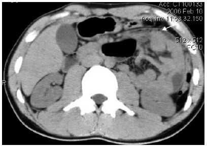
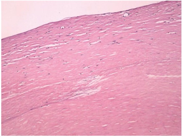
Sclerosing encapsulating peritonitis (SEP) is a rare cause of intestinal obstruction that is characterized by a thick grayish-white fibrotic membrane encasing the small bowel. SEP can be classified as idiopathic, also known as abdominal cocoon, or secondary. It is difficult to make a definite pre-operative diagnosis. We experienced five cases of abdominal cocoon, and the case files were reviewed retrospectively for the clinical presentation, operative findings and outcome. All the patients presented with acute, subacute and chronic intestinal obstruction. Computed tomography (CT) showed characteristic findings of small bowel loops congregated to the center of the abdomen encased by a soft-tissue density mantle in four cases. Four cases had an uneventful post-operative period, one case received second adhesiolysis due to persistent ileus. The imaging techniques may facilitate pre-operative diagnosis. Surgery is important in the management of SEP.
Figures
References
-
- Foo KT, Ng KC, Rauff A, Foong WC, Sinniah R. Unusual small intestinal obstruction in adolescent girls: the abdominal cocoon. Br J Surg. 1978;65:427–430. - PubMed
-
- Sahoo SP, Gangopadhyay AN, Gupta DK, Gopal SC, Sharma SP, Dash RN. Abdominal cocoon in children: a report of four cases. J Pediatr Surg. 1996;31:987–988. - PubMed
-
- Afthentopoulos IE, Passadakis P, Oreopoulos DG, Bargman J. Sclerosing peritonitis in continuous ambulatory peritoneal dialysis patients: one center's experience and review of the literature. Adv Ren Replace Ther. 1998;5:157–167. - PubMed
-
- Holland P. Sclerosing encapsulating peritonitis in chronic ambulatory peritoneal dialysis. Clin Radiol. 1990;41:19–23. - PubMed
Publication types
MeSH terms
LinkOut - more resources
Full Text Sources
Medical
