Review
. 2007 Sep-Oct;92(1-2):43-55.
doi: 10.1016/j.ymgme.2007.06.008. Epub 2007 Jul 30.

A feat of metabolic proportions: Pdx1 orchestrates islet development and function in the maintenance of glucose homeostasis

Affiliations
Review

A feat of metabolic proportions: Pdx1 orchestrates islet development and function in the maintenance of glucose homeostasis

Daniella A Babu et al. Mol Genet Metab. 2007 Sep-Oct.

Abstract

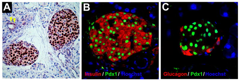
Emerging evidence over the past decade indicates a central role for transcription factors in the embryonic development of pancreatic islets and the consequent maintenance of normal glucose homeostasis. Pancreatic and duodenal homeobox 1 (Pdx1) is the best studied and perhaps most important of these factors. Whereas deletion or inactivating mutations of the Pdx1 gene causes whole pancreas agenesis in both mice and humans, even haploinsufficiency of the gene or alterations in its expression in mature islet cells causes substantial impairments in glucose tolerance and the development of a late-onset form of diabetes known as maturity onset diabetes of the young. The study of Pdx1 has revealed crucial phenotypic interrelationships of the varied cell types within the pancreas, particularly as these impinge upon cellular differentiation in the embryo and neogenesis and regeneration in the adult. In this review, we describe the actions of Pdx1 in the developing and mature pancreas and attempt to unify these actions with its known roles in modulating transcriptional complex formation and chromatin structure at the molecular genetic level.


Figures

Figure 1
Figure 1. Pdx1 expression and gross pancreatic morphology during mouse embryonic development
Shown are schematic representations of the developing mouse pancreas at different embryonic ages. At approximately embryonic day 8.5 (E8.5) signals from the notochord, including fibroblast growth factor 2 (FGF2) and activin, repress expression of hedgehog proteins (HH) in the region of the gut endoderm that is destined to develop into pancreatic buds (dark blue). Initial pancreatic bud outgrowth occurs between E9–9.5 (primary transition), and subsequent branching morphogenesis is dependent upon the expression of Pdx1 (see text). During the secondary transition (between E11–15), there is differentiation of islet cell types (e.g. α and β cells) and acinar cells that is dependent upon the action of Pdx1. After E15, the two buds fuse to form the single organ seen in adult mammals. In adults, Pdx1 expression persists at highest levels in islets (particularly β cells) and at low levels in duct cells and acinar cells.
Figure 2
Figure 2. Pdx1 protein expression in the adult pancreas
Pancreas from a 10 week-old mouse was fixed in paraformaldehyde and embedded in paraffin. Sections were then hematoxylin stained and subject to immunohistochemistry or immunofluorescence. A, horseradish peroxidase-based immunostaining for Pdx1. Pdx1 (dark brown color) demonstrates an islet-specific expression pattern with nuclear localization. Occasional duct cells are also observed to express Pdx1 (yellow arrowheads). B, dual immunofluorescence of an islet using antibodies against Pdx1 (green) and insulin (red). Pdx1 is seen to localize within the nuclei of insulin-producing β cells. C, dual immunofluorescence of an islet using antibodies against Pdx1 (green) and glucagon (red). Pdx1 is not expressed in glucagon-positive α cells. In panels B and C nuclei are counterstained with hoechst H33342 dye (blue).
Figure 3
Figure 3. Upstream regulators and direct downstream targets of Pdx1
The figure emphasizes the central role of Pdx within the pancreas. Cell-extrinsic factors emanating from the notochord (FGF2 and activin) as well as cell-intrinsic factors (transcription factors Foxa2 and HNF6) collaborate to activate Pdx1 expression in the primitive gut endoderm. During pancreas development, Pdx1 is believed to directly activate or repress a subset of genes that lead to differentiation of islet and acinar cells. In the mature pancreas, Pdx1 directly activates or represses genes that confer the β cell phenotype. The “+” sign indicates that the gene is activated by Pdx1, and the “−” sign indicates that the gene is repressed by Pdx1.

References

    1. Kim SK, Hebrok M. Intercellular signals regulating pancreas development and function. Genes Dev. 2001;15:111–127. - PubMed
    1. Scott V, Clark AR, Hutton JC, Docherty K. Two proteins act as the iuf1 insulin gene enhancer binding factor. FEBS Lett. 1991;290:27–30. - PubMed
    1. Ohlsson H, Edlund T. Sequence-specific interactions of nuclear factors with the insulin gene enhancer. Cell. 1986;45:35–44. - PubMed
    1. Ohlsson H, Karlsson O, Edlund T. A beta-cell-specific protein binds to the two major regulatory sequences of the insulin gene enhancer. Proc Natl Acad Sci U S A. 1988;85:4228–4231. - PMC - PubMed
    1. Ohlsson H, Thor S, Edlund T. Novel insulin promoter- and enhancer-binding proteins that discriminate between pancreatic alpha- and beta-cells. Mol Endocrinol. 1991;5:897–904. - PubMed

Publication types