. 2007 Sep;16(9):1830-41.
doi: 10.1110/ps.072883707. Epub 2007 Jul 27.

Sm-like protein Hfq: location of the ATP-binding site and the effect of ATP on Hfq-- RNA complexes

Affiliations

Sm-like protein Hfq: location of the ATP-binding site and the effect of ATP on Hfq-- RNA complexes

Veronique Arluison et al. Protein Sci. 2007 Sep.

Abstract

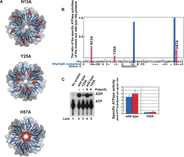
Sm-like proteins are ubiquitous ring-shaped oligomers that exhibit a variety of nucleic acid-binding activities. They have been linked functionally to various cellular events involving RNA, and it is generally believed that their activity is exerted via the passive binding of nucleic acids. Our earlier studies of the Sm-like Escherichia coli protein Hfq provided the first evidence indicating that Hfq is an ATP-binding protein. Using a combination of biochemical and genetic techniques, we have now determined a plausible ATP-binding site in Hfq and tested Hfq's ATP-binding affinity and stoichiometry. The results of RNA footprinting and binding analyses suggest that ATP binding by the Hfq-RNA complex results in its significant destabilization. RNA footprinting indicates deprotection of Hfq-bound RNA tracts in the presence of ATP, suggestive of their release by the protein. The results reported herein broaden the scope of potential in vivo roles for Hfq and other Sm-like proteins.


Figures

Figure 1.
Figure 1.
The effect of site-specific mutations on the efficiency of ATP hydrolysis by purified Hfq proteins. (A) The location of the N13A, Y25A, and H57A mutation sites (shown in red) in the Hfq molecule. Note that the N13A and H57A panels are viewed looking onto the “bottom” of the disc-shaped hexamer (i.e., N-terminal helical face), whereas the Y25A illustration is viewed looking onto the Loop-4 (“L4”) face of the hexamer. (B) For each of the indicated mutations, parallel purifications of the wild-type and mutant Hfq proteins were carried out, and the Hfq-specific ATPase activities were determined as described previously (Sukhodolets and Garges 2003; see page 8023 therein). The ratios of the two specific activities are plotted versus the Hfq amino acid sequence. Data represent the average of two or more independent experiments. (Blue columns) Previously constructed mutations (Sukhodolets and Garges 2003); (red columns) mutations described in this study. (C) A representative PEI-cellulose plate on which the activities of the Hfq near-knockout Y25A mutant protein and the wild-type Hfq protein are tested side by side. The quantitated results of this experiment are shown at right; reactions were performed in the (blue columns) absence or in the (red columns) presence of excess Poly(rA). Data represent the average of two independent experiments.
Figure 2.
Figure 2.
The effect of ATP on Hfq–RNA interaction. Experiments using 5′(fluorescein)-modified RNA probes were carried out as described in Materials and Methods. (A) ATP diminishes the RNA-binding activity of Hfq. Titrations of rA7 in the (rectangles) absence or in the (triangles) presence of 1 mM AMP-PNP, (rhombuses) 1 μM ATP, or (circles) 1 mM ATP with wild-type Hfq produced K d values of 27, 28, 30, and 100 nM, respectively. The decrease in fluorescence anisotropy in the presence of excess ATP is consistent with our central claim that ATP modulates RNA–Hfq interactions; we believe that the anisotropy change may reflect complex interactions between RNA-fluorophore and the Hfq hexamer in an altered conformation. (B) Titration of rA7 with Y25A Hfq resulted in the determination of a K d value of 118 nM.
Figure 3.
Figure 3.
Determination of the number of ATP-binding sites and the affinities of Hfq for nucleotides. Experiments using MANT-modified nucleotides were carried out as described in Materials and Methods. (A) Titration of MANT-ATP with the Hfq protein. The equivalence point, which was reached at ∼10 μM Hfq monomer (at 10 μM MANT-ATP), gives a binding stoichiometry of 6 ATP molecules per Hfq hexamer. (B) Titrations of (rectangles) MANT-ATP, (triangles) MANT-ADP, and (circles) MANT-AMP-PNP with Hfq produced similar dissociation constants of ∼0.75 μM. (C) Parallel titrations of MANT-ATP with (rectangles) wild-type and (triangles) Y25A Hfq gave dissociation constants of ∼0.75 and 1.75 μM, respectively.
Figure 4.
Figure 4.
ATP destabilizes Hfq–RNA complexes. (A) Schematic diagram of the secondary structure of the RNA probe used in these experiments. Key Ribonuclease A cleavage sites are indicated by arrows. (B) Effect of ATP on Hfq binding to a model RNA incorporating a stem–loop structure and 3′-terminal poly(rA) tail studied by Ribonuclease A footprinting. RNA footprinting experiments were carried out as described in Materials and Methods. Reactions in the absence or in the presence of 1 mM ATP-γ-S are shown. The de-protected sites are indicated by arrowheads. (C) Destabilization of the urea-insensitive Hfq–RNA complexes in the presence of ATP. EMSA experiments were carried out as described in Materials and Methods.
Figure 5.
Figure 5.
Disruption of the Hfq sixfold symmetry in the RNA–Hfq–ATP complex. The electron microscopic experiments were carried out as described in Materials and Methods. The lack of the effect in the ternary complexes using non-hydrolyzable ATP analogs suggests that the conformational change accompanying this transition is coupled to ATP hydrolysis.
Figure 6.
Figure 6.
Functional effects of the Hfq Y25A mutation. Coupled transcription–translation in hfq-minus S30 extracts in the presence of (black columns) 0.2 μM purified wild-type Hfq, (gray columns) 0.2 μM Hfq Y25A, or (open columns) in the absence of Hfq. The coupled in vitro transcription–translation reactions used a pBESTluc™ supercoiled DNA template and were carried out as described (Sukhodolets and Garges 2003).
Figure 7.
Figure 7.
Structural model for the Hfq ATP-binding site. A. The Hfq hexamer is viewed onto the “L4” face, with protein chains represented as ribbon cartoons. Semitransparent solvent-accessible surfaces are drawn in alternating gray intensities, and an individual Hfq monomer is delimited by dashed lines (β-strands and termini are indicated for this subunit). ATP (green) and Y25 (tan) are rendered as space-filling CPK spheres, and the ATP-binding cleft is shown in more detail in B. Surface colors for residues within 4.5 Å of ATP are graded from (yellow) least conserved to (orange) moderately conserved and (red) most strictly conserved amino acids. (C) In addition to favorable π···π stacking interactions, numerous other ionic and hydrogen-bond contacts are indicated, with the width of the dashed lines (magenta) that represent the interaction scaled by the degree of phylogenetic conservation of that amino acid. The phosphoester tail for alternative, less energetically favorable configurations of bound ATP are drawn as thin lines. Note that the binding pocket lies along the boundary between individual Hfq subunits, and that strongly conserved residues contribute to form a composite binding site capable of providing a network of Hfq···ATP interactions to staple the nucleotide into this surface pocket.

Similar articles

Cited by

References

    1. Arluison V., Derreumaux, P., Allemand, F., Folichon, M., Hajnsdorf, E., and Régnier, P. 2002. Structural modeling of the Sm-like protein Hfq from Escherichia coli . J. Mol. Biol. 320 705–712. - PubMed
    1. Arluison V., Mura, C., Guzman, M.R., Liquier, J., Pellegrini, O., Gingery, M., Regnier, P., and Marco, S. 2006. Three-dimensional structures of fibrillar Sm proteins: Hfq and other Sm-like proteins. J. Mol. Biol. 356 86–96. - PubMed
    1. Azam T.A., Hiraga, S., and Ishihama, A. 2000. Two types of localization of the DNA-binding proteins within the Escherichia coli nucleoid. Genes Cells 5 613–626. - PubMed
    1. Brescia C.C., Mikulecky, P.J., Feig, A.L., and Sledjeski, D.D. 2003. Identification of the Hfq-binding site on DsrA RNA: Hfq binds without altering DsrA secondary structure. RNA 9 33–43. - PMC - PubMed
    1. DuBow M.S., Ryan, T., Young, R.A., and Blumenthal, T. 1977. Host factor for coliphage Q β RNA replication: Presence in procaryotes and association with the 30S ribosomal subunit in Escherichia coli . Mol. Gen. Genet. 153 39–43. - PubMed

Publication types

MeSH terms

LinkOut - more resources