. 2007 Sep;16(9):1984-98.
doi: 10.1110/ps.072773007. Epub 2007 Jul 27.

Molecular plasticity of beta-catenin: new insights from single-molecule measurements and MD simulation

Affiliations

Molecular plasticity of beta-catenin: new insights from single-molecule measurements and MD simulation

Monica Ritco-Vonsovici et al. Protein Sci. 2007 Sep.

Abstract

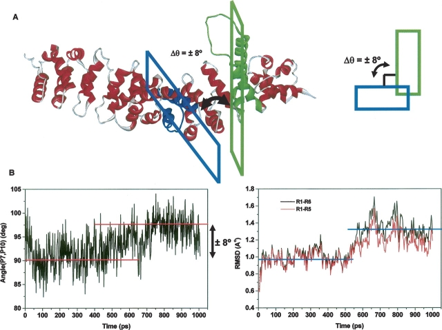
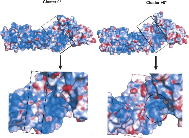
The multifunctional protein, beta-catenin, has essential roles in cell adhesion and, through the Wnt signaling pathway, in controlling cell differentiation, development, and generation of cancer. Could distinct molecular forms of beta-catenin underlie these two functions? Our single-molecule force spectroscopy of armadillo beta-catenin, with molecular dynamics (MD) simulation, suggests a model in which the cell generates various forms of beta-catenin, in equilibrium. We find beta-catenin and the transcriptional factor Tcf4 form two complexes with different affinities. Specific cellular response is achieved by the ligand binding to a particular matching preexisting conformer. Our MD simulation indicates that complexes derive from two conformers of the core region of the protein, whose preexisting molecular forms could arise from small variations in flexible regions of the beta-catenin main binding site. This mechanism for the generation of the various forms offers a route to tailoring future therapeutic strategies.


Figures

Figure 1.
Figure 1.
Tcf4/armadillo β-catenin recognition. Purified armadillo β-catenin (residues 134–671) was allowed to interact either with GST-Tcf4 (residues 1–57) (A,B) or His-Tcf4 (C,D). The interaction in A and B was detected with goat anti-GST antibody, HRP-anti-goat IgG, and HRP substrate. (A) Purified armadillo β-catenin was adsorbed to ELISA plates. (B) Purified armadillo β-catenin was specifically attached to a mica substrate via flexible linkers. (C) In the AFM molecular recognition setup, His–Tcf4 was specifically attached to the cantilever tip via PEG linkers and brought into contact with armadillo β-catenin attached to the mica substrate via EGS linkers. (D) Raw data from a representative force–distance cycle measured at 0.5 Hz for armadillo β-catenin/Tcf4 interaction. The functionalized tip was approached to the functionalized surface and then retracted while the deflection of the cantilever was monitored. The attractive force signal in the retrace curve reflects the unbinding event, its magnitude corresponding to the unbinding force, fu.
Figure 2.
Figure 2.
Distributions of unbinding forces (A–E) and force spectra (F) for armadillo β-catenin/Tcf4 complexes. Histograms from 200–400 approach/retract cycles of the complex are presented at various probe velocities: (A) 40 nm/sec, (B) 100 nm/sec, (C) 200 nm/sec, (D) 1160 nm/sec, and (E) 2330 nm/sec. The maximum of each distribution was found by the Gaussian fit of the histograms. Two maxima are evident for most of the distributions, corresponding to the presence of two populations of molecules. Increasing the probe velocity and the loading rate results in the shift in the peak distributions to higher forces and the increase of the fractional occupancy of the low-strength population. (F) Most probable unbinding forces were plotted against the logarithm of the loading rate. Two plots are presented corresponding to the two populations of bound armadillo β-catenin/Tcf4 complexes.
Figure 3.
Figure 3.
Global analysis of MD trajectory of apo-armadillo β-catenin. (A) Ribbon representation of the minimized apo structure with the long loop of R10 (green) modeled. (B) Backbone RMSD of all repeats with and without the R10 loop (top left), the radius of gyration of all repeats (top right), backbone RMSD of R7 and R10 without the loop (bottom left), and backbone atom fluctuations of all repeats (bottom right).
Figure 4.
Figure 4.
Angle analysis within and adjacent to R7 and R10 of apo-armadillo β-catenin during the MD trajectory. From top to bottom and for either R7 (left) or R10 (right): time-dependent torsion angle between H2 and H3 helices, torsion angle between helices H−1 and H2, and torsion angle between helices H3 and H+1. The events are marked by arrows.
Figure 5.
Figure 5.
Existence of two molecular populations of apo-armadillo β-catenin. (A) Ribbon diagram of apo-armadillo β-catenin highlighting the perpendicular arrangement between planes formed by H2 and H3 helices within R7 and R10 (P7 and P10), which may change by 8°, resulting in two molecular populations. (B) Time-dependent angle change between P7 and P10 planes (left) and time-dependent RMSD of either repeats 1–6 or repeats 1–5 (right).
Figure 6.
Figure 6.
Statistical distribution and force spectra of unbinding forces for the armadillo β-catenin/Tcf4 interaction in the presence of ICAT. Histograms from 200–300 approach/retract cycles of the complex at 2330 nm/sec are presented. One maximum is evident for these distributions, corresponding to the presence of one population of molecules. In the absence of ICAT (A) the low-strength complex is populated but in the presence of ICAT (B) the high-strength population 2 is favored. (C) Four force spectra plots are presented corresponding to the two populations of bound armadillo β-catenin/Tcf4 complexes in the presence (▾,★, dashed lines) or absence of ICAT (▪, •, solid lines).
Figure 7.
Figure 7.
Electrostatic potential surfaces of representative structures from MD simulation of apo-armadillo β-catenin. The electrostatic potential surfaces (EPS) of representative structure from MD simulation at 0° rotation (Cluster-0) and at 8° rotation (Cluster-8) were colored blue for positive and red for negative EPS, respectively. Upper figure represents the EPS of the whole structure and lower figure the zoom on the major groove of armadillo β-catenin.

Similar articles

Cited by

References

    1. Behrens J. and Lustig, B. 2004. The Wnt connection to tumorigenesis. Int. J. Dev. Biol. 48 477–487. - PubMed
    1. Behrens J., von Kries, J.P., Kuhl, M., Bruhn, L., Wedlich, D., Grosschedl, R., and Birchmeier, W. 1996. Functional interaction of β-catenin with the transcription factor LEF-1. Nature 382 638–642. - PubMed
    1. Case D.L., Darden, T.A., Cheatham, T.E., Simmerling, C.L., Wang, J., Duke, R.E., Luo, R., Merz, K.M., Wang, B., Pearlman, D.A., et al. 2004. AMBER 8. University of California, San Francisco, CA.
    1. Changeux J.P. and Edelstein, S.J. 2005. Allosteric mechanisms of signal transduction. Science 308 1424–1428. - PubMed
    1. Choi H.J., Huber, A.H., and Weis, W.I. 2006. Thermodynamics of β-catenin–ligand interactions: The roles of the N- and C-terminal tails in modulating binding affinity. J. Biol. Chem. 281 1027–1038. - PubMed

Publication types

MeSH terms