. 2007 Aug 21;104(34):13603-8.
doi: 10.1073/pnas.0702756104. Epub 2007 Jul 27.

Recombinant human butyrylcholinesterase from milk of transgenic animals to protect against organophosphate poisoning

Affiliations

Recombinant human butyrylcholinesterase from milk of transgenic animals to protect against organophosphate poisoning

Yue-Jin Huang et al. Proc Natl Acad Sci U S A. .

Abstract

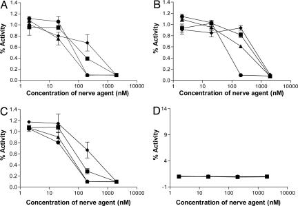
Dangerous organophosphorus (OP) compounds have been used as insecticides in agriculture and in chemical warfare. Because exposure to OP could create a danger for humans in the future, butyrylcholinesterase (BChE) has been developed for prophylaxis to these chemicals. Because it is impractical to obtain sufficient quantities of plasma BChE to treat humans exposed to OP agents, the production of recombinant BChE (rBChE) in milk of transgenic animals was investigated. Transgenic mice and goats were generated with human BChE cDNA under control of the goat beta-casein promoter. Milk from transgenic animals contained 0.1-5 g/liter of active rBChE. The plasma half-life of PEGylated, goat-derived, purified rBChE in guinea pigs was 7-fold longer than non-PEGylated dimers. The rBChE from transgenic mice was inhibited by nerve agents at a 1:1 molar ratio. Transgenic goats produced active rBChE in milk sufficient for prophylaxis of humans at risk for exposure to OP agents.


Conflict of interest statement

The authors declare no conflict of interest.

Figures

Fig. 1.
Fig. 1.
Generation of rBChE transgenic animals. (A) Schematic of the bCN-BChE transgene used to generate the transgenic animals. The lower bars indicate the lengths of the PCR products and the primer pairs used for DIG-labeling of probes and screening transgenic animals. (B) Southern blot analysis of HindIII-digested genomic mouse DNA. Lane 1, DIG-labeled Marker III; lanes 2–8, genomic DNA from transgenic mice in the following order: 61-2B3F, 43-3A11F, 43-3A13F, 43-3A14F, 43-3A14A2F, 43-3A15F, and 43-3A15A9F; lane 9, genomic DNA from a nontransgenic FVB mouse; lanes 10–14, serial dilutions (2×, 4×, 8×, 16×, and 32×) of transgene plasmid DNA spiked into genomic DNA of a nontransgenic FVB mouse. The membrane was hybridized with Acb712-244. The arrow indicates the rBChE transgene (≈6.2 kb). (C) Southern blot analysis of SacI-SphI digested genomic goat DNA. Lane 1, DIG-labeled Marker III; lanes 2–7, genomic DNA from transgenic goats 1871F, 1876M, 1880F, 1900F, 1902F, and 1925F; lane 8, genomic DNA from a nontransgenic goat; lanes 9–12, serial dilutions (3×, 6×, 12×, and 24×) of transgene plasmid DNA spiked into genomic DNA of a nontransgenic goat. The membrane was hybridized with Acb712-244. The arrow indicates the rBChE transgene (≈6.5 kb). Endogenous BChE showed a weak band of ≈15 kb. (D) FISH analysis of metaphase spreads from a skin fibroblast cell sample of an F2 transgenic goat, with an insulator probe. Arrow indicates localization of the rBChE transgene detected by FITC (shown in green) on a goat chromosome. Chromosomes were stained with DAPI (shown in blue).
Fig. 2.
Fig. 2.
rBChE expression, activity, and oligomeric forms in the milk of transgenic animals from natural lactations. For PAGE, 10 μl of samples were loaded onto each lane of a precast 4–20% Tris-glycine gel unless otherwise noted. (A) BChE activity gel staining of milk samples from transgenic mice. Lane 1, purified plasma huBChE (25 units/ml); lane 2, diluted 61-2B3F milk (1:50); lane 3, diluted 43-3A-15F milk (1:1,000); lane 4, diluted 44-2 milk (1:3); lane 5, diluted FVB mouse milk (1:3). (B) BChE activity gel staining of milk samples from transgenic goats. Lane 1, purified plasma huBChE (10 units/ml); lane 2, diluted milk from a nontransgenic goat (1:2); lanes 3–5, diluted milk containing rBChE from 3 transgenic goats: 1871F (1:80), 2220F (1:40), and 5719F (1:40). (C) Western blot analysis of milk from transgenic mice under denaturing and reducing conditions. Lane 1, purified plasma huBChE (25 units/ml); lane 2, diluted 61-2B3F milk (1:25); lane 3, diluted 43-3A-15F milk (1:500, 30 μl); lane 4, diluted 44-2 milk (1:1.5, 20 μl); lane 5, diluted FVB mouse milk (1:1.5). (D) Western blot analysis of milk from transgenic goats under denaturing and reducing conditions. Lane 1, purified plasma huBChE (10 units/ml); lane 2, diluted milk from a nontransgenic goat (1:2); lanes 3–5, diluted milk containing rBChE from 3 transgenic goats: 1871F (1:80), 2220F (1:40), and 5719F (1:40). (E) Analysis of milk from a transgenic goat by SEC-HPLC. Raw milk samples from 5719F, an F2 goat of 2219M, were briefly centrifuged then loaded into a SEC-HPLC system. Fractions were collected over time and analyzed by the Ellman assay. The specific activity of the rBChE versus SEC-HPLC collection intervals was plotted. (F) Silver-stained SDS/PAGE on purified rBChE from the milk of transgenic goats. Lane 1, diluted milk from a nontransgenic goat (1:100); lane 2, diluted milk from the transgenic founder goat, 1871F (1:100); lane 3, diluted milk from the transgenic goat, 2220F (1:100); lane 4, purified plasma huBChE (10 units/ml); lane 5, purified rBChE from milk of the transgenic founder goat, 1871F (250 ng); lane 6, purified rBChE from milk of the transgenic goat, 2220F (250 ng).
Fig. 3.
Fig. 3.
Clearance of purified rBChE by guinea pigs. The guinea pigs were injected IV (A) or IM (B) with PEGylated rBChE (▴) or dimer rBChE (■). Residual BChE activity (units/ml), measured in blood by the Ellman assay, was plotted over time (hours).
Fig. 4.
Fig. 4.
In vitro inhibition by nerve agents of rBChE contained in the milk of transgenic mice. Binding was performed with diluted raw mouse milk samples from three transgenic mice [3A14F (A), 3A15F (B), and 2B3F (C)] and a control FVB mouse (D). The reactions were carried out in the presence of tabun (GA, ♦), sarin (GB, ■), soman (GD, ▴), and VX (●), respectively. Residual enzyme activity was measured by the Ellman assay. Data points represent the mean ± SD from duplicates in each mouse.

Comment in

References

    1. Lockridge O, Bartels CF, Vaughan TA, Wong CK, Norton SE, Johnson LL. J Biol Chem. 1987;262:549–557. - PubMed
    1. Ostergaard D, Viby-Mogensen J, Hanel HK, Skovgaard LT. Acta Anaesthesiol Scand. 1988;32:266–269. - PubMed
    1. Cerasoli DM, Griffiths EM, Doctor BP, Saxena A, Fedorko JM, Greig NH, Yu QS, Huang Y, Wilgus H, Karatzas CN, et al. Chem Biol Interact. 2005;157–158:363–365. - PubMed
    1. Lenz DE, Yeung D, Smith JR, Sweeney RE, Lumley LA, Cerasoli DM. Toxicology. 2007;233:31–39. - PubMed
    1. Darvesh S, Hopkins DA, Geula C. Nat Rev Neurosci. 2003;4:131–138. - PubMed

Publication types

MeSH terms