Novel tonoplast transporters identified using a proteomic approach with vacuoles isolated from cauliflower buds
- PMID: 17660356
- PMCID: PMC1976570
- DOI: 10.1104/pp.107.096917
Novel tonoplast transporters identified using a proteomic approach with vacuoles isolated from cauliflower buds
Abstract
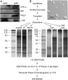
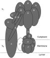
Young meristematic plant cells contain a large number of small vacuoles, while the largest part of the vacuome in mature cells is composed by a large central vacuole, occupying 80% to 90% of the cell volume. Thus far, only a limited number of vacuolar membrane proteins have been identified and characterized. The proteomic approach is a powerful tool to identify new vacuolar membrane proteins. To analyze vacuoles from growing tissues we isolated vacuoles from cauliflower (Brassica oleracea) buds, which are constituted by a large amount of small cells but also contain cells in expansion as well as fully expanded cells. Here we show that using purified cauliflower vacuoles and different extraction procedures such as saline, NaOH, acetone, and chloroform/methanol and analyzing the data against the Arabidopsis (Arabidopsis thaliana) database 102 cauliflower integral proteins and 214 peripheral proteins could be identified. The vacuolar pyrophosphatase was the most prominent protein. From the 102 identified proteins 45 proteins were already described. Nine of these, corresponding to 46% of peptides detected, are known vacuolar proteins. We identified 57 proteins (55.9%) containing at least one membrane spanning domain with unknown subcellular localization. A comparison of the newly identified proteins with expression profiles from in silico data revealed that most of them are highly expressed in young, developing tissues. To verify whether the newly identified proteins were indeed localized in the vacuole we constructed and expressed green fluorescence protein fusion proteins for five putative vacuolar membrane proteins exhibiting three to 11 transmembrane domains. Four of them, a putative organic cation transporter, a nodulin N21 family protein, a membrane protein of unknown function, and a senescence related membrane protein were localized in the vacuolar membrane, while a white-brown ATP-binding cassette transporter homolog was shown to reside in the plasma membrane. These results demonstrate that proteomic analysis of highly purified vacuoles from specific tissues allows the identification of new vacuolar proteins and provides an additional view of tonoplastic proteins.
Figures


Similar articles
-
Identification of a vacuolar sucrose transporter in barley and Arabidopsis mesophyll cells by a tonoplast proteomic approach.Plant Physiol. 2006 May;141(1):196-207. doi: 10.1104/pp.106.079533. Epub 2006 Mar 31. Plant Physiol. 2006. PMID: 16581873 Free PMC article.
-
A proteomics dissection of Arabidopsis thaliana vacuoles isolated from cell culture.Mol Cell Proteomics. 2007 Mar;6(3):394-412. doi: 10.1074/mcp.M600250-MCP200. Epub 2006 Dec 6. Mol Cell Proteomics. 2007. PMID: 17151019 Free PMC article.
-
iTRAQ-based quantitative proteomic analysis reveals the role of the tonoplast in fruit senescence.J Proteomics. 2016 Sep 2;146:80-9. doi: 10.1016/j.jprot.2016.06.031. Epub 2016 Jun 28. J Proteomics. 2016. PMID: 27371350
-
Vacuolar transporters and their essential role in plant metabolism.J Exp Bot. 2007;58(1):83-102. doi: 10.1093/jxb/erl183. Epub 2006 Nov 16. J Exp Bot. 2007. PMID: 17110589 Review.
-
Vacuolar Transporters - Companions on a Longtime Journey.Plant Physiol. 2018 Feb;176(2):1384-1407. doi: 10.1104/pp.17.01481. Epub 2018 Jan 2. Plant Physiol. 2018. PMID: 29295940 Free PMC article. Review.
Cited by
-
Glutathione and glutathione-S-transferase activities of the vacuoles of the beet (Beta vulgaris L.) roots.Dokl Biol Sci. 2010 Jul-Aug;433:275-8. doi: 10.1134/S0012496610040113. Epub 2010 Aug 17. Dokl Biol Sci. 2010. PMID: 20711876 No abstract available.
-
GABA Metabolism, Transport and Their Roles and Mechanisms in the Regulation of Abiotic Stress (Hypoxia, Salt, Drought) Resistance in Plants.Metabolites. 2023 Feb 26;13(3):347. doi: 10.3390/metabo13030347. Metabolites. 2023. PMID: 36984787 Free PMC article. Review.
-
Elemental profiling and genome-wide association studies reveal genomic variants modulating ionomic composition in Populus trichocarpa leaves.Front Plant Sci. 2024 Nov 28;15:1450646. doi: 10.3389/fpls.2024.1450646. eCollection 2024. Front Plant Sci. 2024. PMID: 39670268 Free PMC article.
-
A Review of Plant Vacuoles: Formation, Located Proteins, and Functions.Plants (Basel). 2019 Sep 5;8(9):327. doi: 10.3390/plants8090327. Plants (Basel). 2019. PMID: 31491897 Free PMC article. Review.
-
Functional and physiological characterization of Arabidopsis INOSITOL TRANSPORTER1, a novel tonoplast-localized transporter for myo-inositol.Plant Cell. 2008 Apr;20(4):1073-87. doi: 10.1105/tpc.107.055632. Epub 2008 Apr 25. Plant Cell. 2008. PMID: 18441213 Free PMC article.
References
-
- Apse MP, Aharon GS, Snedden WA, Blumwald E (1999) Salt tolerance conferred by overexpression of a vacuolar Na+/H+ antiport in Arabidopsis. Science 285 1256–1258 - PubMed
-
- Barrieu F, Thomas D, Marty-Mazars D, Charbonnier M, Marty F (1998) Tonoplast intrinsic proteins from cauliflower (Brassica oleracea L. var. botrytis): immunological analysis, cDNA cloning and evidence for expression in meristematic tissues. Planta 204 335–344 - PubMed
-
- Bednarczyk D, Ekins S, Wikel JH, Wright SH (2003) Influence of molecular structure on substrate binding to the human organic cation transporter, hOCT1. Mol Pharmacol 63 489–498 - PubMed
-
- Bradford MM (1976) Rapid and quantitative method for quantitation of microgram quantities of protein utilizing the principle of protein-dye binding. Anal Biochem 72 248–252 - PubMed
Publication types
MeSH terms
Substances
LinkOut - more resources
Full Text Sources
Molecular Biology Databases

