. 2007 Oct;6(10):1841-52.
doi: 10.1128/EC.00205-07. Epub 2007 Jul 27.

FEA1, FEA2, and FRE1, encoding two homologous secreted proteins and a candidate ferrireductase, are expressed coordinately with FOX1 and FTR1 in iron-deficient Chlamydomonas reinhardtii

Affiliations

FEA1, FEA2, and FRE1, encoding two homologous secreted proteins and a candidate ferrireductase, are expressed coordinately with FOX1 and FTR1 in iron-deficient Chlamydomonas reinhardtii

Michael D Allen et al. Eukaryot Cell. 2007 Oct.

Abstract

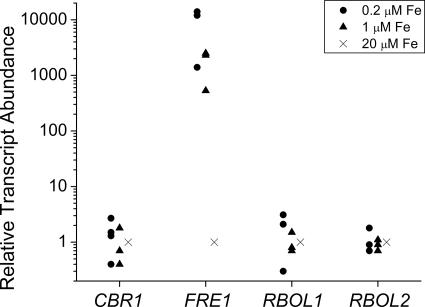
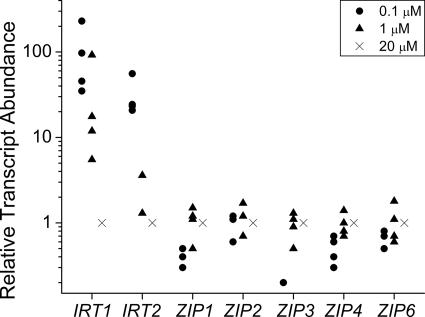
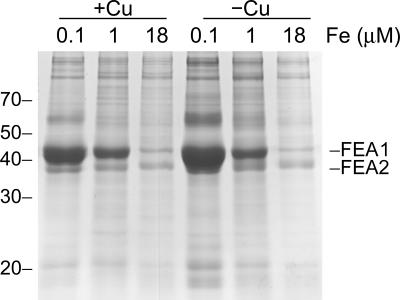
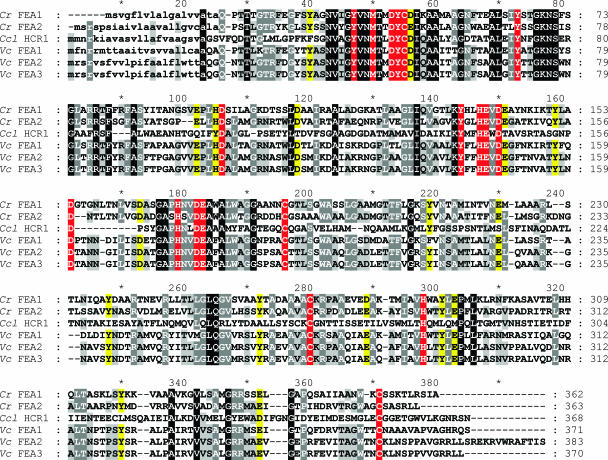
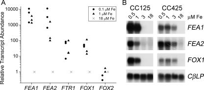
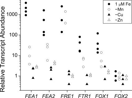
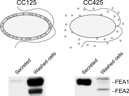
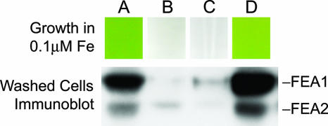
Previously, we had identified FOX1 and FTR1 as iron deficiency-inducible components of a high-affinity copper-dependent iron uptake pathway in Chlamydomonas. In this work, we survey the version 3.0 draft genome to identify a ferrireductase, FRE1, and two ZIP family proteins, IRT1 and IRT2, as candidate ferrous transporters based on their increased expression in iron-deficient versus iron-replete cells. In a parallel proteomic approach, we identified FEA1 and FEA2 as the major proteins secreted by iron-deficient Chlamydomonas reinhardtii. The recovery of FEA1 and FEA2 from the medium of Chlamydomonas strain CC425 cultures is strictly correlated with iron nutrition status, and the accumulation of the corresponding mRNAs parallels that of the Chlamydomonas FOX1 and FTR1 mRNAs, although the magnitude of regulation is more dramatic for the FEA genes. Like the FOX1 and FTR1 genes, the FEA genes do not respond to copper, zinc, or manganese deficiency. The 5' flanking untranscribed sequences from the FEA1, FTR1, and FOX1 genes confer iron deficiency-dependent expression of ARS2, suggesting that the iron assimilation pathway is under transcriptional control by iron nutrition. Genetic analysis suggests that the secreted proteins FEA1 and FEA2 facilitate high-affinity iron uptake, perhaps by concentrating iron in the vicinity of the cell. Homologues of FEA1 and FRE1 were identified previously as high-CO(2)-responsive genes, HCR1 and HCR2, in Chlorococcum littorale, suggesting that components of the iron assimilation pathway are responsive to carbon nutrition. These iron response components are placed in a proposed iron assimilation pathway for Chlamydomonas.


Figures

FIG. 1.
FIG. 1.
Transcripts for one of four candidate ferrireductases are increased in iron-deficient medium. Candidate genes were analyzed by quantitative real-time reverse transcription-PCR for expression during iron-deficient growth. Strains 17D, CC1021, and CC125 were tested on 0.2, 1, or 20 μM iron. Each 20-μl reaction mixture contained cDNA equivalent to 100 ng of input total mRNA. Fold induction was calculated according to the 2−ΔΔCT method (50). Each data point is the average of a technical triplicate and represents an individual experiment.
FIG. 2.
FIG. 2.
Survey of candidate metal transporter genes for expression in iron-deficient medium. Candidate genes for transporters were analyzed by real time reverse transcription-PCR for expression in strains CC125 and CC1021 grown in 0.1, 1, or 20 μM iron. Relative transcript abundance was calculated as described in the legend to Fig. 1.
FIG. 3.
FIG. 3.
FEA1 and FEA2 are secreted by iron-deficient cells. Proteins secreted into the medium by a Chlamydomonas cw15 strain (CC400) were collected by ammonium sulfate/trichloroacetic acid precipitation and analyzed after electrophoretic separation on SDS-containing polyacrylamide (12% monomer) gels. Each lane contained extract corresponding to an equivalent number (7.3 × 10⁷) of cells. The major proteins revealed by Coomassie blue staining were analyzed by MALDI-TOF mass spectrometry after in-gel trypsin digestion.
FIG. 4.
FIG. 4.
Conserved residues in the FEA proteins. FEA1 (BAA94959) and FEA2 (protein ID 173281 in the version three draft genome) of C. reinhardtii (Cr), Hcr1 (BAA22844) of Chlorococcum littorale (Ccl), and C_370140 (Fea1), C_520002 (Fea2), and C_520026 (Fea3) of V. carteri (Vc) (version 1.0 draft genome) were aligned using Multalin (http://prodes.toulouse.inra.fr/multalin/multalin.html) (11). Signal peptides, indicated by lowercase letters, and cleavage sites were predicted using TargetP (http://www.cbs.dtu.dk/services/TargetP/) (24, 59). All are predicted to be secreted proteins. Residues are indicated by white text on a black background if identical in all six sequences or on a gray background if identical in five of the six or by black text on a gray background if four of six are conserved. Potential metal binding ligands are indicated with a red background if they are identical or with a yellow background if the residues represent conservative replacements. Asterisks mark every 10th residue.
FIG. 5.
FIG. 5.
FEA1 and FEA2 are expressed coordinately with FOX1 and FTR1 genes under iron-deficient conditions. (A) Real-time PCR quantitation of gene expression in CC125. See the legend to Fig. 1. Each data point is the average of a technical triplicate and represents an individual experiment. (B) Blot hybridization. Wild-type (CC125) or wall-less (CC425) cells were grown in TAP medium with iron supplementation as indicated. RNA was isolated after 5 days of growth, corresponding to cell densities in the range of 5 × 10⁶ to 7 × 10⁶ cell/ml. Five micrograms of total RNA was loaded in each lane and analyzed by blot hybridization. The expression of CBLP was monitored as a loading control.
FIG. 6.
FIG. 6.
Metal-selective response of FEA gene expression. Transcript abundance was assessed by real-time PCR (see the legend to Fig. 1). For copper and manganese deficiency, cells were adapted to deficiency by three sequential transfers (1:1,000 dilution at each transfer) into medium lacking the trace element, while for zinc deficiency, cells were sampled after the second transfer into zinc-free medium. For iron deficiency, the cells were washed once and transferred from iron-replete (18 μM) to iron-deficient (1 μM) medium and sampled when they reached a density of 5 × 10⁶ to 7 × 10⁶ cell/ml. The various RNAs collected from strains CC125, CC425, and CC1021 were tested for marker gene expression characteristic for each deficiency (CYC6 for copper deficiency, GPX1 for manganese deficiency, and ZRT1 for zinc deficiency). For each experiment, the level of expression is normalized to the replete condition. Each data point is the average of a technical triplicate and represents an individual experiment.
FIG. 7.
FIG. 7.
Functional model for FEA proteins. Top, FEA proteins (circles) associate within the periplasm in a cell wall-containing Chlamydomonas strain. In a cell wall-less mutant, FEA proteins are lost to the medium and unable to function in iron assimilation. Bottom, secreted and cell-associated fractions of CC125 (walled) and CC425 (wall-deficient) cells were isolated and separated by denaturing gel electrophoresis. FEA proteins were visualized by immunoblotting. Each lane corresponds to ∼2 × 10⁶ cells.
FIG. 8.
FIG. 8.
Loss of FEA proteins results in poor growth on low-iron medium. Spores from a cw15 arg2 mt+ × CW15 ARG2 mt− tetrad were analyzed for growth on iron-limited (0.1 μM) TAP-Arg medium (top) and cell-associated FEA proteins by immunoblot analysis (bottom) of washed iron-deficient cells (grown on TAP-Arg with 1 μM Fe).
FIG. 9.
FIG. 9.
Model for iron assimilation by Chlamydomonas. The components of an iron assimilation pathway are shown. Ferric iron may be released from chelated forms by reduction to the ferrous form. The FEA proteins may bind the ferrous iron to keep it soluble and available as a substrate for the ferroxidase/ferric transporter complex. Alternatively, FEA proteins might bind ferric ion, in which case they would function upstream of FRE1. FRE1 was found in a plasma membrane subproteome (70), which is consistent with the model proposed here.

Similar articles

Cited by

References

    1. Abboud, S., and D. J. Haile. 2000. A novel mammalian iron-regulated protein involved in intracellular iron metabolism. J. Biol. Chem. 275:19906-19912. - PubMed
    1. Allen, M. D., J. Kropat, S. Tottey, J. A. Del Campo, and S. S. Merchant. 2007. Manganese deficiency in Chlamydomonas results in loss of photosystem II and MnSOD function, sensitivity to peroxides, and secondary phosphorus and iron deficiency. Plant Physiol. 143:263-277. - PMC - PubMed
    1. Askwith, C., D. Eide, A. Van Ho, P. S. Bernard, L. Li, S. Davis-Kaplan, D. M. Sipe, and J. Kaplan. 1994. The FET3 gene of S. cerevisiae encodes a multicopper oxidase required for ferrous iron uptake. Cell 76:403-410. - PubMed
    1. Askwith, C., and J. Kaplan. 1997. An oxidase-permease-based iron transport system in Schizosaccharomyces pombe and its expression in Saccharomyces cerevisiae. J. Biol. Chem. 272:401-405. - PubMed
    1. Bagnaresi, P., S. Thoiron, M. Mansion, M. Rossignol, P. Pupillo, and J. F. Briat. 1999. Cloning and characterization of a maize cytochrome-b5 reductase with Fe3+-chelate reduction capability. Biochem. J. 338:499-505. - PMC - PubMed

Publication types

MeSH terms

LinkOut - more resources