. 2007 Sep;177(1):281-93.
doi: 10.1534/genetics.107.076133. Epub 2007 Jul 29.

Cdc15 is required for spore morphogenesis independently of Cdc14 in Saccharomyces cerevisiae

Affiliations

Cdc15 is required for spore morphogenesis independently of Cdc14 in Saccharomyces cerevisiae

M Evangelina Pablo-Hernando et al. Genetics. 2007 Sep.

Abstract

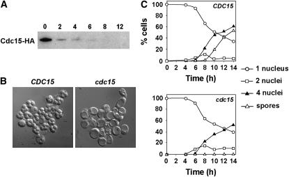
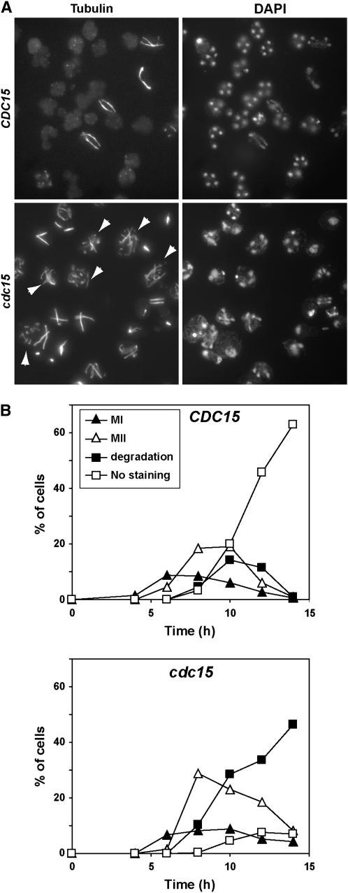
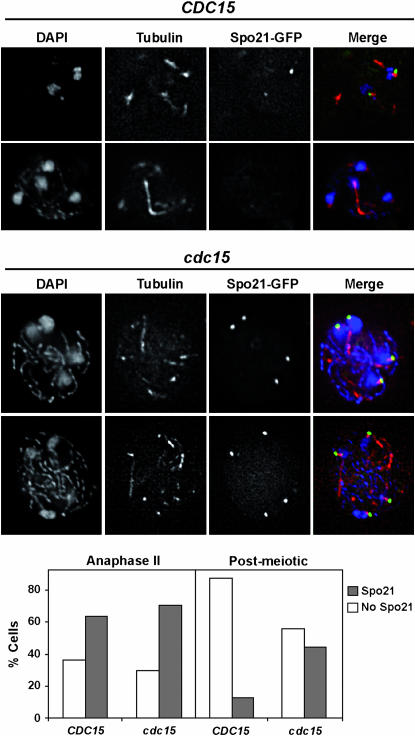
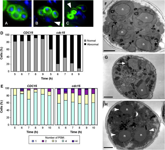
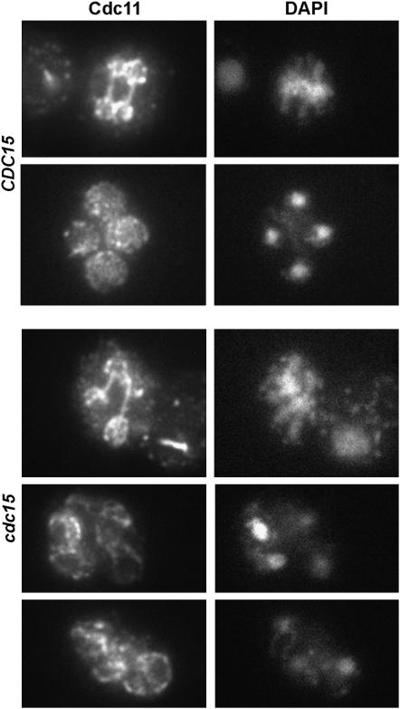
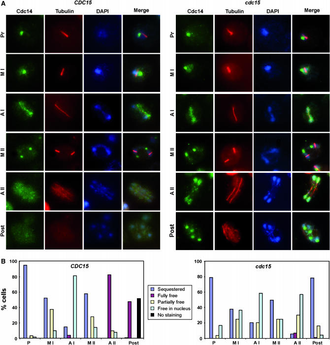
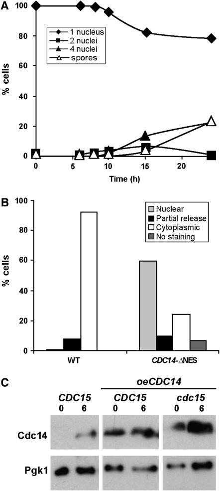
In Saccharomyces cerevisiae exit from mitosis requires the Cdc14 phosphatase to reverse CDK-mediated phosphorylation. Cdc14 is released from the nucleolus by the Cdc14 early anaphase release (FEAR) and mitotic exit network (MEN) pathways. In meiosis, the FEAR pathway is essential for exit from anaphase I. The MEN component Cdc15 is required for the formation of mature spores. To analyze the role of Cdc15 during sporulation, a conditional mutant in which CDC15 expression was controlled by the CLB2 promoter was used. Cdc15-depleted cells proceeded normally through the meiotic divisions but were unable to properly disassemble meiosis II spindles. The morphology of the prospore membrane was aberrant and failed to capture the nuclear lobes. Cdc15 was not required for Cdc14 release from the nucleoli, but it was essential to maintain Cdc14 released and for its nucleo-cytoplasmic transport. However, cells carrying a CDC14 allele with defects in nuclear export (Cdc14-DeltaNES) were able to disassemble the spindle and to complete spore formation, suggesting that the Cdc14 nuclear export defect was not the cause of the phenotypes observed in cdc15 mutants.


Figures

F<sc>igure</sc> 1.—
Figure 1.—
CDC15 is not required for DNA replication or meiotic segregation. (A) Levels of Cdc15 protein in cells with the PCLB2-CDC15 construct. Cdc15 was immunoprecipitated from 1 mg of protein extracts before Western blot analysis. Antibodies against the N-terminal HA epitope tags were used for detection and immunoprecipitation. Samples were collected at the indicated number of hours after transfer to sporulation medium (0, 2, 4, 6, 8, and 12 hr). (B) Depletion of CDC15 during sporulation blocks the formation of spores. DIC images of wild type (CDC15) or PCLB2-CDC15 mutant (cdc15) incubated for 24 hr in sporulation medium. (C) Time course of appearance of binucleate cells (squares) or tetranucleate cells (triangles) in wild type (CDC15) or PCLB2-CDC15 mutant (cdc15). Strains were incubated in sporulation medium for the indicated times (hours) before aliquots were collected and the cells were stained with DAPI. At least 200 cells were counted for each time point.
F<sc>igure</sc> 2.—
Figure 2.—
Cdc15-depleted cells have defects in meiotic spindle disassembly. (A) Nuclear and spindle morphology in wild-type and cdc15 mutants. Wild type (CDC15) and PCLB2-CDC15 mutant (cdc15) were incubated in sporulation medium; samples were collected at different time intervals and stained with anti-tubulin antibody (left) and DAPI (right). The image shows an example of cells that had been incubated in sporulation medium for 10 hr. (B) Quantification of the different spindle morphologies during the sporulation process in wild-type (CDC15) and PCLB2-CDC15 (cdc15) cells. Cells from the meiotic time course shown in Figure 1C were stained with anti-tubulin antibody and classified according to the spindle morphology. Meiosis I includes cells in prophase, metaphase I, and anaphase I, whereas cells with metaphase II or anaphase II spindles were classified as meiosis II. Postmeiotic cells that had already disassembled the meiotic spindle and were negative for tubulin staining were classified as “no staining.” Cells in meiosis II with broken or collapsed spindles were classified as “degradation.”
F<sc>igure</sc> 3.—
Figure 3.—
cdc15 mutants have defects in SPB modification. Spo21 localizes to the SPB in Cdc15-depleted cells during sporulation. Wild type (CDC15, top) or PCLB2-CDC15 mutant (cdc15, bottom) expressing SPO21-GFP (from plasmid pSPO21) were transferred to sporulation medium. After 8 hr of incubation, cells were collected and stained with anti-tubulin antibody and DAPI. Images of Spo21-GFP (green), tubulin (red), and DAPI (blue) and the merging of the three channels are shown. The graph shows the percentage of wild-type (CDC15) or cdc15 mutant cells with Spo21 staining (open rectangles) or without staining (shaded rectangles) in anaphase II or postmeiotic cells. At least 100 cells were counted for each category.
F<sc>igure</sc> 4.—
Figure 4.—
PCLB2-CDC15 cells display PSM defects. Fluorescence micrographs of sporulating wild-type (A) and PCLB2-CDC15 cells (B and C). PSMs are visualized in green using GFP-Spo2051−91, and nuclei are shown in blue using DAPI. Arrowheads in B indicate unpackaged nuclei in postmeiotic cells and abnormal PSMs in C. (D) Quantification of the PSM morphology during sporulation in wild type (CDC15) and PCLB2-CDC15 mutant (cdc15). At least 200 cells in which the PSM was visible were counted for each strain. The graph shows the percentage of cells with normal morphology (shaded bars) or aberrant shapes (solid bars) in cells with visible PSMs. Cells with at least one abnormal PSM were counted as aberrant. (E) Quantification of the number of PSMs during sporulation in wild type (CDC15) and PCLB2-CDC15 mutant (cdc15). At least 200 cells in which the PSM was visible were counted for each strain. (F–H) Electron micrographs of sporulating wild type (F) and PCLB2-CDC15 cells (G and H). White arrow in G indicates a growing PSM that is not encapsulating a nucleus (indicated with n in all images). Arrowheads in H indicate multiple, unpackaged nuclei in a postmeiotic cell. Bars, 1 μm.
F<sc>igure</sc> 5.—
Figure 5.—
Septins are normally assembled in cdc15 mutants. Localization of Cdc11 in wild type and PCLB2-CDC15 mutant. Cells were incubated in sporulation medium. Samples were collected at different times and stained with anti-Cdc11 antibody and DAPI and inspected for septin morphology. Images of Cdc11 (left) and DAPI (right) are shown.
F<sc>igure</sc> 6.—
Figure 6.—
Localization of Cdc14 during sporulation. (A) Wild type (CDC15, left) or PCLB2-CDC15 mutant (cdc15, right) were transferred to sporulation medium. At different times after the induction of sporulation, cells were collected and stained with anti-Cdc14 and anti-tubulin antibodies and DAPI. Anti-goat Alexa Fluor 488 and anti-mouse Alexa Fluor 633 were used as secondary antibodies. Cdc14 (green), tubulin (red), and DAPI (blue) images are shown, as well as the merged image. Representative images of the different meiotic phases are shown for each strain. (B) Localization of Cdc14 during the different stages of the sporulation in wild-type (left) or PCLB2-CDC15 (right) cells. Localization of Cdc14 in each phase of the sporulation process was classified as sequestered in the nucleolus (blue), fully (maroon) or partially (yellow) released from the nucleolus, free in the nucleus (light blue), or cells showing no staining (black). For wild-type cells, the different localization is presented as a percentage of metaphase I (MI; n = 109), anaphase I (AI; n = 92), metaphase II (MII; n = 149), anaphase II (AII; n = 155), or spores (n = 113) with visible Cdc14. For PCLB2-CDC15 cells, a similar percentage for metaphase I (MI; n = 112), anaphase I (AI; n = 74), metaphase II (MII; n = 128), anaphase II (AII; n = 132), or postmeiotic cells (n = 64) is shown.
F<sc>igure</sc> 7.—
Figure 7.—
Importance of Cdc14 nuclear export in sporulation. Cdc14-ΔNES strains have defects in sporulation. Time course of appearance of binucleate (squares) and tetranucleate cells (triangles) and spores (white triangles) in cells carrying Cdc14-ΔNES (SEP41). Cells were incubated in sporulation medium for the indicated times (hours) before aliquots were collected and stained with DAPI. At least 200 cells were counted for each time point. (B) Localization of Cdc14 and Cdc14-ΔNES in anaphase II cells. Cells were collected and stained with anti-Cdc14 and anti-tubulin antibodies and DAPI. Localization of Cdc14 in the fraction of cells that had entered meiosis was classified as nuclear (lightly shaded bar), cytoplasmic (open bars), partially released (solid bars), or without staining (darkly shaded bar). At least 100 cells in anaphase II were counted. (C) Analysis of Cdc14 protein levels in the wild-type strain (CDC15) and in the wild-type and cdc15 mutant strains overexpressing CDC14 (CDC15 oeCDC14 and cdc15 oeCDC14, respectively). Samples were collected at 0 and 6 hr after transfer to sporulation medium to prepare protein extracts. Western blot was performed using anti-Cdc14 antibody. Anti-Pgk1 was used as loading control.

Similar articles

Cited by

References

    1. Asakawa, K., and A. Toh-e, 2002. A defect of Kap104 alleviates the requirement of mitotic exit network gene functions in Saccharomyces cerevisiae. Genetics 162: 1545–1556. - PMC - PubMed
    1. Bajgier, B. K., M. Malzone, M. Nickas and A. M. Neiman, 2001. SPO21 is required for meiosis-specific modification of the spindle pole body in yeast. Mol. Biol. Cell 12: 1611–1621. - PMC - PubMed
    1. Bardin, A. J., and A. Amon, 2001. MEN and SIN: What's the difference? Nat. Rev. Mol. Cell. Biol. 2: 815–826. - PubMed
    1. Bembenek, J., J. Kang, C. Kurischko, B. Li, J. R. Raab et al., 2005. Crm1-mediated nuclear export of Cdc14 is required for the completion of cytokinesis in budding yeast. Cell Cycle 4: 961–971. - PubMed
    1. Buonomo, S. B., K. P. Rabitsch, J. Fuchs, S. Gruber, M. Sullivan et al., 2003. Division of the nucleolus and its release of CDC14 during anaphase of meiosis I depends on separase, SPO12, and SLK19. Dev. Cell. 4: 727–739. - PubMed

Publication types

MeSH terms