

doi: 10.1534/genetics.107.075283.
Epub 2007 Jul 29.
Natural variation of ebony gene controlling thoracic pigmentation in Drosophila melanogaster
Affiliations
- PMID: 17660557
- PMCID: PMC2034628
- DOI: 10.1534/genetics.107.075283
Item in Clipboard
Natural variation of ebony gene controlling thoracic pigmentation in Drosophila melanogaster
Genetics.
2007 Oct.
Abstract
We identified the causal genetic variation for the difference in the thoracic trident pigmentation intensity between two wild-derived strains of Drosophila melanogaster. It was found to be the difference in expression level of ebony, which codes for an enzyme in the melanin-synthesis pathway and has pleiotropic effects on vision and behavior.
Figures
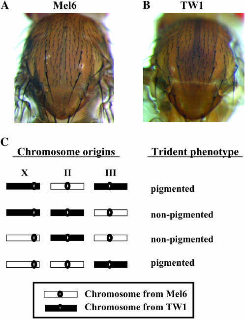
Thoracic trident pigmentation pattern and its association with the third chromosome. (A) Thorax segment of Mel6 (G59) originated from Benin, West Africa. (B) Thorax segment of TW1 (G23) originated from Taiwan. Mel6 (G59) and TW1 (G23) were subjected to 23 and 59 generations of sib mating, respectively, to construct nearly isogenic lines. (C) Trident phenotypes of the whole-chromosome substitution lines between Mel6 and TW1. Chromosome origins and the trident phenotypes of the lines are indicated in the left and right columns, respectively. These lines were generated using balancer chromosomes by a similar scheme used in Hollocher et al. (1997). Pigmentation intensity was examined under the microscope.
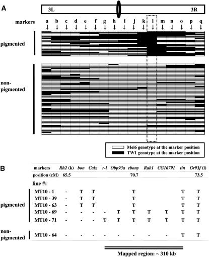
Genetic mapping of the thoracic trident pigmentation phenotype using 73 homozygous recombinant lines constructed between TW1 and Mel6. (A) Genotypes of the pigmented (n = 19) and nonpigmented (n = 54) phenotype lines at 17 restriction fragment length polymorphism (RFLP) markers (a–q) on the third chromosome: a, Gr61d; b, gry; c, Gr64A; d, lama; e, Gr65c; f, Dhpr; g, Gr68d; h, Or74a; i, desat2; j, pnr; k, Rh2; l, Gr93f; m, RpS3; n, ssh; o, Pr; p, Gr98a; q, Ptx. Horizontal bars indicate genotypes of the recombinant lines. The dotted square indicates the genotypes at Gr93f marker that had almost complete association with the phenotype (r2 = 0.93). Note that one exceptional line that showed nonpigmented phenotype but had TW1 genotype at Gr93f is represented as MT10-64 in B. RFLP markers were developed by direct sequencing PCR fragments of ∼600–900 bp from Mel6 and TW1. Further information on RFLP markers can be provided upon request. Primers were designed by Primer3 software (Rozen and Skaletsky 2000) from the publicly available genome sequence (Adams et al. 2000). DNA was extracted from one male each from Mel6 and TW1 using the GeneElute Mammalian Genomic DNA kit (Sigma-Aldrich, St. Louis). PCR products were obtained from these genomic samples by the AmpliTaq Gold PCR kit (Applied Biosystems, Foster City, CA) and were purified using MultiScreen-FB filter plates (Millipore, Billerica, MA). These purified products were directly sequenced on both strands using the BigDye terminator cycle sequencing kit version 3 (Applied Biosystems). The third chromosome recombinant lines were constructed from F2 backcrossed individuals in the Mel6 homozygous background. The line was backcrossed twice (including the F2 backcross) to Mel6 and had two generations of recombination opportunity. The lines were made homozygous using balancers in the last step. Note that these third chromosome recombinants possess the X and the second chromosomes from Mel6. (B) Genotypes at eight additional RFLP markers in six recombinant lines from A that had their breakpoints between markers Rh2 (k) and Gr93f (l). T indicates TW1 genotype and “-” indicates Mel6 genotype. Two critical lines, MT10-69 and MT10-64, indicate that the ∼310-kb region between markers r-l and tin is responsible for the phenotype.
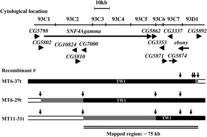
Recombination breakpoints of the three most informative recombinants between Mel6 and TW1. The recombinants were screened from a laboratory population that experienced over 20 generations of recombination. These third chromosome recombinants had the X and the second chromosomes from Mel6. Horizontal arrows indicate annotated ORFs with their transcriptional directions. Vertical arrowheads indicate positions of the markers genotyped. Open and solid bars represent chromosome segments from Mel6 and TW1, respectively. Shaded bars indicate regions where recombinant breakpoints reside. The markers were genotyped by directly sequencing PCR fragments of ∼600–900 bp. The thoracic tridents of the three recombinants were all pigmented. Note that the critical interval of ∼75kb between CG7000 and CG5892 includes the candidate gene ebony.
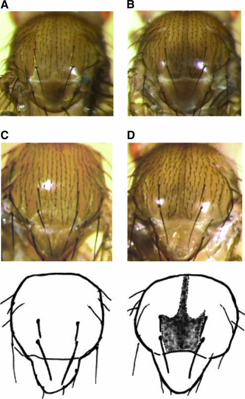
Thoracic trident pigmentation phenotype obtained from complementation test with ebony and ebony RNAi knockdown experiment. (A) Dorsal view of the thorax segment of the Mel6/e1 heterozygote. (B) Dorsal view of the thorax segment of the TW1/e1 heterozygote. The flies in A and B were reared at 25°. The results were almost identical when eAFA, Df(3R)e-H4, and Df(3R)e-R1 were used as ebony mutants (data not shown). (C) Dorsal view of the thorax segment of the wild-type control, w1118; P{w+mc = Actin5C-GAL4}/+. (D) Dorsal view of the thorax segment of the RNAi knockdown individual, w1118; P{UAS-ebony-IR}/+; P{w+mc = Actin5C-GAL4}/+. The insert, P{UAS-ebony-IR} contains two fragments of 500 bp ebony cDNA as an inverted repeat (IR). These lines were crossed to w1118; P{w+mc = Act5C–GAL4}17bFO1/+ strain to induce IR RNA in the whole body. w1118; P{w+mc = Act5C–GAL4}17bFO1/+ was obtained by backcrossing y1 w*; P{w+mc = Act5C–GAL4}17bFO1/TM6B, Tb1 to w1118 strain for 10 generations. Adult flies carrying one copy of the UAS-IR construct and one copy of the GAL4 driver were investigated for the thorax pigmentation. The flies in C and D were reared at 20°.
Similar articles
-
Divergent enhancer haplotype of ebony on inversion In(3R)Payne associated with pigmentation variation in a tropical population of Drosophila melanogaster.Mol Ecol. 2011 Oct;20(20):4277-87. doi: 10.1111/j.1365-294X.2011.05260.x. Epub 2011 Sep 13. Mol Ecol. 2011. PMID: 21914015
-
Complex patterns of cis-regulatory polymorphisms in ebony underlie standing pigmentation variation in Drosophila melanogaster.Mol Ecol. 2015 Dec;24(23):5829-41. doi: 10.1111/mec.13432. Epub 2015 Nov 20. Mol Ecol. 2015. PMID: 26503353
-
Pigmentation and behavior: potential association through pleiotropic genes in Drosophila.Genes Genet Syst. 2013;88(3):165-74. doi: 10.1266/ggs.88.165. Genes Genet Syst. 2013. PMID: 24025245 Review.
-
The molecular genetics of clinal variation: a case study of ebony and thoracic trident pigmentation in Drosophila melanogaster from eastern Australia.Mol Ecol. 2011 May;20(10):2100-10. doi: 10.1111/j.1365-294X.2011.05089.x. Epub 2011 Apr 5. Mol Ecol. 2011. PMID: 21466604
-
The Genetic Basis of Pigmentation Differences Within and Between Drosophila Species.Curr Top Dev Biol. 2016;119:27-61. doi: 10.1016/bs.ctdb.2016.03.004. Epub 2016 Apr 25. Curr Top Dev Biol. 2016. PMID: 27282023 Free PMC article. Review.
Cited by
-
Genetic basis of sex-specific color pattern variation in Drosophila malerkotliana.Genetics. 2008 Sep;180(1):421-9. doi: 10.1534/genetics.108.091728. Epub 2008 Aug 24. Genetics. 2008. PMID: 18723880 Free PMC article.
-
Adaptation to local ultraviolet radiation conditions among neighbouring Daphnia populations.Proc Biol Sci. 2011 May 7;278(1710):1306-13. doi: 10.1098/rspb.2010.1663. Epub 2010 Oct 13. Proc Biol Sci. 2011. PMID: 20943691 Free PMC article.
-
Transcriptome analysis reveals novel patterning and pigmentation genes underlying Heliconius butterfly wing pattern variation.BMC Genomics. 2012 Jun 29;13:288. doi: 10.1186/1471-2164-13-288. BMC Genomics. 2012. PMID: 22747837 Free PMC article.
-
Unraveling the Tangled Skein: The Evolution of Transcriptional Regulatory Networks in Development.Annu Rev Genomics Hum Genet. 2015;16:103-31. doi: 10.1146/annurev-genom-091212-153423. Epub 2015 May 20. Annu Rev Genomics Hum Genet. 2015. PMID: 26079281 Free PMC article. Review.
-
Reconciling Differences in Pool-GWAS Between Populations: A Case Study of Female Abdominal Pigmentation in Drosophila melanogaster.Genetics. 2016 Feb;202(2):843-55. doi: 10.1534/genetics.115.183376. Epub 2015 Dec 29. Genetics. 2016. PMID: 26715669 Free PMC article.
References
-
- Adams, M. D., S. E. Celniker, R. A. Holt, C. A. Evans, J. D. Gocayne et al., 2000. The genome sequence of Drosophila melanogaster. Science 287: 2185–2195. - PubMed
-
- Gibert, P., B. Moreteau and J. R. David, 2000. Developmental constraints on an adaptive plasticity: reaction norms of pigmentation in adult segments of Drosophila melanogaster. Evol. Dev. 2: 249–260. - PubMed
-
- Gibert, P., P. Capy, A. Imasheva, B. Moreteau, J. P. Morin et al., 2004. Comparative analysis of morphological traits among Drosophila melanogaster and D. simulans: genetic variability, clines and phenotypic plasticity. Genetica 120: 165–179. - PubMed
Publication types
MeSH terms
Substances
Associated data
- Actions
- Actions
LinkOut - more resources
Full Text Sources
Molecular Biology Databases
