. 2007 Oct;55(13):1385-96.
doi: 10.1002/glia.20544.

IFN-gamma-induced IDO and WRS expression in microglia is differentially regulated by IL-4

Affiliations

IFN-gamma-induced IDO and WRS expression in microglia is differentially regulated by IL-4

Manisha C Yadav et al. Glia. 2007 Oct.

Abstract

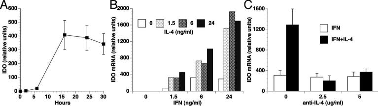
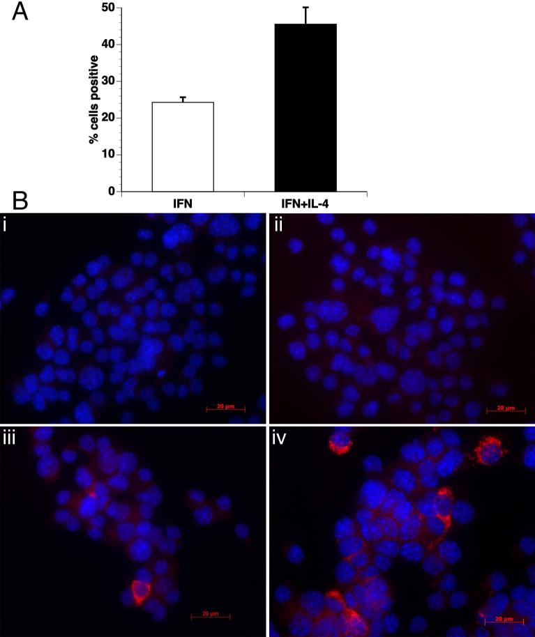
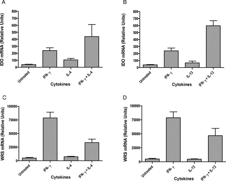
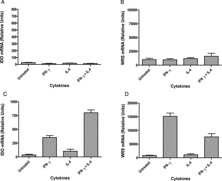
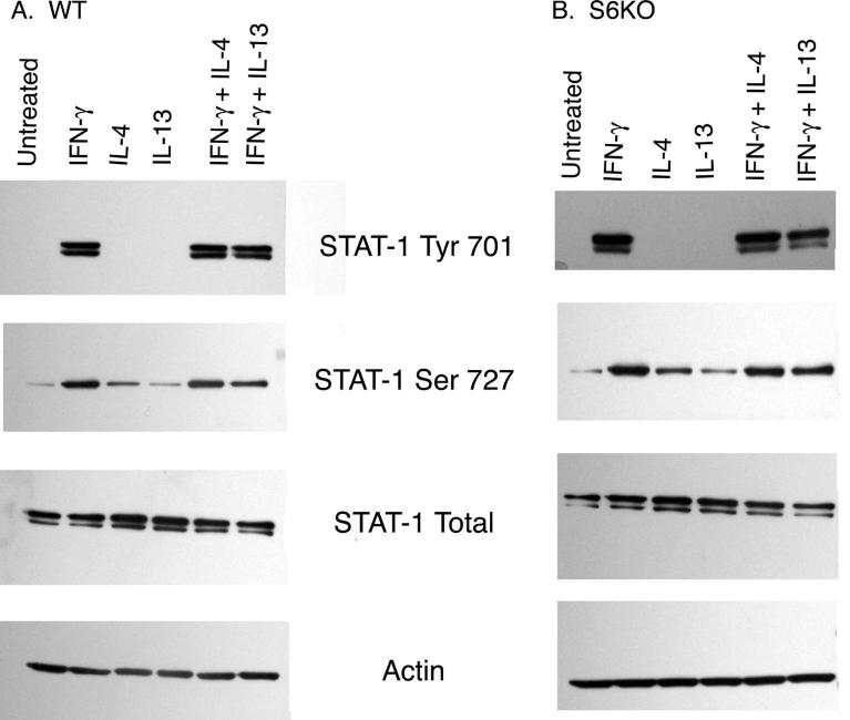
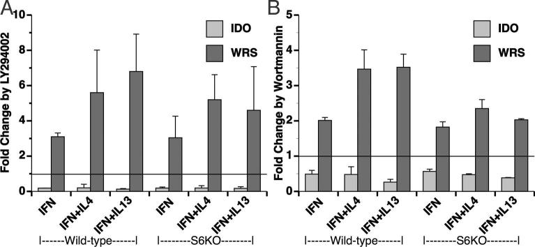
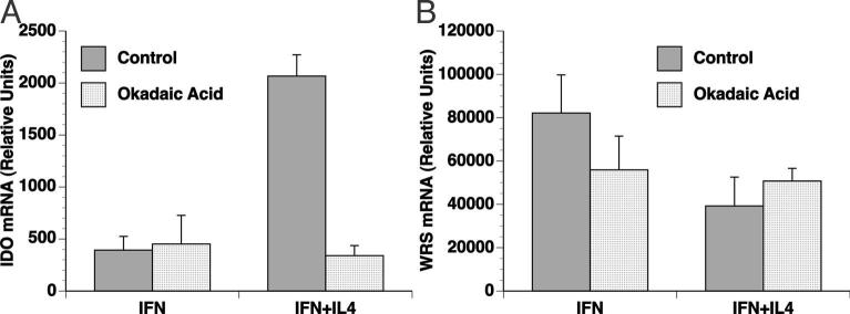
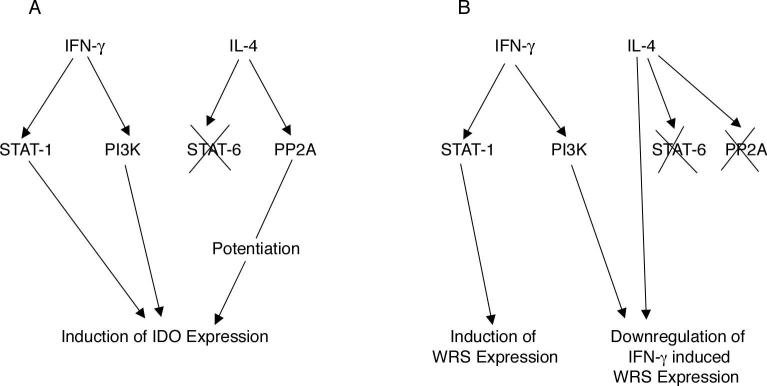
Indoleamine 2,3-dioxygenase (IDO), a tryptophan catabolizing enzyme, has been implicated in the pathogenesis of various neurological disorders. IDO expression is induced by IFN-gamma and leads to neurotoxicity by generating quinolinic acid. Additionally, it inhibits the immune response through both tryptophan depletion and generating other tryptophan catabolites. IL-4 and IL-13 have been shown to control IDO expression by antagonizing the effects of IFN-gamma in different cell types. Here, we investigated the effects of these cytokines on IDO expression in microglia. Interestingly, we observed that both IL-4 and IL-13 greatly enhanced IFN-gamma-induced IDO expression. However, tryptophanyl-tRNA synthetase (WRS), which is coinduced with IDO by IFN-gamma, is downregulated by IL-4 and IL-13. The effect of IL-4 and IL-13 was independent of STAT-6. Modulation of IDO but not WRS was eliminated by inhibition of protein phosphatase 2A (PP2A) activity. The phosphatidylinositol 3-kinase (PI3K) pathway further differentiated the regulation of these two enzymes, as inhibiting the PI3K pathway eliminated IFN-gamma induction of IDO, whereas such inhibition greatly enhanced WRS expression. These findings show discordance between modulations of expression of two distinct enzymes utilizing tryptophan as a common substrate, and raise the possibility of their involvement in regulating immune responses in various neurological disorders.


Figures

Figure 1
Figure 1. IL-4 increases IFN-γ -induced IDO mRNA expression
(A) EOC13.31 microglia cells were treated with 10 ng/ml IFN-γ for the indicated time periods, and total RNA was analyzed for IDO expression by real time RT-PCR. (B) EOC13.31 microglia cells were treated with the indicated concentrations of IFN-γ and IL-4 for 24 hours, and total RNA was analyzed for IDO expression by real time RT-PCR. (C) EOC13.31 microglia cells were treated with 10 ng/ml of IFN-γ with and without 25 ng/ml of IL-4 for 24 h, in the presence of the indicated concentration of anti-IL-4 antisera. Total RNA was analyzed for IDO expression by real time RT-PCR.
Figure 2
Figure 2. IL-4 increases IFN-γ-induced IDO protein expression
Cellular expression of the IDO protein was examined by immunofluorescence using anti-IDO antibody in EOC13.31 cells. Cells were either untreated, or treated with IFN-γ, IL-4 or IFN-γ+IL-4. (A) Increased numbers of cells had detectable IDO when treated with IFN-γ + IL-4 as compared to IFN-γ alone. Mean and SEM of 5 different fields, data are representative of two independent experiments. (B) Untreated (i) and cells treated with IL-4 alone (ii) did not show any positive staining for IDO. IDO was present in cells treated with IFN-γ alone (iii), and increased in cells treated with IFN-γ+IL-4 (iv).
Figure 3
Figure 3. Isolation and purity of primary microglia
Microglia were isolated from a day-10 mixed culture (obtained from brains of 1-2 days old pups) by magnetic column separation using CD11b beads. The proportion of microglial cells was routinely ∼95% based on detection with FACS staining using anti CD11b antibody. Percentage of cells stained with CD11b before column separation shown in red grey and after column separation shown in blue black (A). Purity was also checked by immunocytochemistry (B) using DAPI nuclear counter stain (i), anti-Iba-1 antibody for microglia (ii) and anti-GFAP antibody for astrocytes (iii). Juxtaposed image of cells stained with DAPI, Iba-1 and GFAP (iv).
Figure 4
Figure 4. IL-4 and IL-13 enhance IFN-γ -induced IDO expression but reduce WRS expression in mouse microglia.
Microglia were stimulated with cytokines for 24 hrs and total RNA was analyzed for IDO (A, B) and WRS (C, D) expression by real time RT-PCR. Data are representative of six independent experiments with IL-4 and seven independent experiments with IL-13 done in triplicate. IDO expression in IFN-γ stimulated cells was significantly increased by treatment with IL-4 or IL-13, whereas IFN-γ stimulated WRS expression significantly reduced by IL-4 or IL-13.
Figure 5
Figure 5. IFN-γ utilizes STAT-1 and IL-4 act independent of STAT-6 in modulating IDO and WRS expression.
Microglia from S1KO (A, B) and S6KO (C, D) mice were stimulated with cytokines for 24 hrs and total RNA was analyzed for IDO and WRS expression by real time RT-PCR. Results are representative of three independent experiments for S1KO and six independent experiments for S6KO done in triplicate. In S6KO mice, IL-4 still significantly increased IDO and decreases WRS in IFN-γ stimulated cells.
Figure 6
Figure 6. IL-4 and IL-13 do not act by altering phosphorylation status of STAT-1
Microglia from WT (A) and S6KO (B) mice were stimulated with cytokines for 30 min, lysed, and subjected to Western blot analysis using anti total STAT-1, anti pSTAT-1 Tyr701, anti pSTAT-1 Ser727 and anti-actin (as a protein loading control). Blots shown are the representative of one of the three independent experiments.
Figure 7
Figure 7. Inhibition of PI3 Kinase reduces IDO but enhances WRS expression.
Microglia from WT and S6KO mice were pretreated with the PI3K inhibitors 20 μM LY (A) or 2 μM wortmannin (WM) (B) for 1 h, followed by cytokine treatments as indicated. Total RNA for IDO and WRS expression was analyzed by real time RT-PCR. Data are normalized to that obtained from cells treated with the drug vehicle (DMSO) only, and represent the mean and SEM of three experiments.
Figure 8
Figure 8. IL-4 potentiation of IFN-γinduced IDO expression utilizes a protein phosphatase.
Microglia from WT mice were pretreated with PP2A inhibitor, 50 nM Okadaic acid (OA) for 1 h, followed by cytokine treatment as indicated. Total RNA for IDO (A) and WRS (B) expression was analyzed by real time RT-PCR. Data are mean and SEM of one representative experiment from three independent experiments. OA significantly reduced IDO induction by IFN-γ+IL-4 but not IFN-γ alone, whereas WRS expression did not significantly differ.
Figure 9
Figure 9. Mechanistic overview of differential modulation of IDO (A) and WRS (B) by IL-4 along with IFN-γ in mouse microglia.
Binding of IFN-γ to its receptor leads to activation of STAT-1, which in turn leads to transcription of IDO and WRS genes. IL-4 potentiates IDO expression but downregulates WRS expression in the presence of IFN-γ in a STAT-6 independent manner. PP2A does not play a role in WRS expression by IFN-γ and/or IL-4, however inhibition of PP2A abrogates IL-4 upregulation of IFN-γ induced IDO expression. IFN-γ also utilizes PI3K pathway in inducing IDO expression as shown by LY and WM (PI3K inhibitors). However, use of LY and WM enhance WRS expression unlike suppression of IDO expression again showing differential regulation of IDO and WRS, two enzymes utilizing tryptophan as a common substrate.

References

    1. Ahmed NN, Grimes HL, Bellacosa A, Chan TO, Tsichlis PN. Transduction of interleukin-2 antiapoptotic and proliferative signals via Akt protein kinase. Proc Natl Acad Sci U S A. 1997;94(8):3627–32. - PMC - PubMed
    1. Bach EA, Aguet M, Schreiber RD. The IFN gamma receptor: a paradigm for cytokine receptor signaling. Annu Rev Immunol. 1997;15:563–91. - PubMed
    1. Bara H, Hainfellner JA, Kepplinger B, Mazal PR, Schmid H, Budka H. Kynurenic acid metabolism in the brain of HIV-1 infected patients. J Neural Transm. 2000;107(10):1127–38. - PubMed
    1. Bialojan C, Takai A. Inhibitory effect of a marine-sponge toxin, okadaic acid, on protein phosphatases. Specificity and kinetics. Biochem J. 1988;256(1):283–90. - PMC - PubMed
    1. Boasso A, Herbeuval JP, Hardy AW, Winkler C, Shearer GM. Regulation of indoleamine 2,3-dioxygenase and tryptophanyl-tRNA-synthetase by CTLA-4-Fc in human CD4+ T cells. Blood. 2005;105(4):1574–81. - PubMed

Publication types

MeSH terms