. 2007 Jul 27:8:56.
doi: 10.1186/1471-2202-8-56.

Sodium channel Nav1.6 accumulates at the site of infraorbital nerve injury

Affiliations

Sodium channel Nav1.6 accumulates at the site of infraorbital nerve injury

Michael A Henry et al. BMC Neurosci. .

Abstract

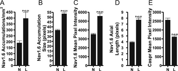
Background: Sodium channel (NaCh) expressions change following nerve and inflammatory lesions and this change may contribute to the activation of pain pathways. In a previous study we found a dramatic increase in the size and density of NaCh accumulations, and a remodeling of NaChs at intact and altered myelinated sites at a location just proximal to a combined partial axotomy and chromic suture lesion of the rat infraorbital nerve (ION) with the use of an antibody that identifies all NaCh isoforms. Here we evaluate the contribution of the major nodal NaCh isoform, Nav1.6, to this remodeling of NaChs following the same lesion. Sections of the ION from normal and ION lesioned subjects were double-stained with antibodies against Nav1.6 and caspr (contactin-associated protein; a paranodal protein to identify nodes of Ranvier) and then z-series of optically sectioned images were captured with a confocal microscope. ImageJ (NIH) software was used to quantify the average size and density of Nav1.6 accumulations, while additional single fiber analyses measured the axial length of the nodal gap, and the immunofluorescence intensity of Nav1.6 in nodes and of caspr in the paranodal region.

Results: The findings showed a significant increase in the average size and density of Nav1.6 accumulations in lesioned IONs when compared to normal IONs. The results of the single fiber analyses in caspr-identified typical nodes showed an increased axial length of the nodal gap, an increased immunofluorescence intensity of nodal Nav1.6 and a decreased immunofluorescence intensity of paranodal caspr in lesioned IONs when compared to normal IONs. In the lesioned IONs, Nav1.6 accumulations were also seen in association with altered caspr-relationships, such as heminodes.

Conclusion: The results of the present study identify Nav1.6 as one isoform involved in the augmentation and remodeling of NaChs at nodal sites following a combined partial axotomy and chromic suture ION lesion. The augmentation of Nav1.6 may result from an alteration in axon-Schwann cell signaling mechanisms as suggested by changes in caspr expression. The changes identified in this study suggest that the participation of Nav1.6 should be considered when examining changes in the excitability of myelinated axons in neuropathic pain models.


Figures

Figure 1
Figure 1
Nav1.6 and caspr staining. A and B. Confocal micrographs from average-intensity z-projections (45 slices in A; 47 slices in B) demonstrate Nav1.6 (red) and caspr (green) staining relationships seen within the normal (A) and lesioned (B) ION. A. The normal ION shows bright Nav1.6 staining (arrowhead) at nodes of Ranvier that are identified as the gap seen between the paranodal staining of caspr. The caspr staining is intense within the paranodal region and prominent Nav1.6 staining is generally restricted to the narrow area at the node. B. The lesioned ION, at a location just proximal to the chromic suture, shows a change in the Nav1.6 and caspr staining relationships that were seen in A. These changes include a loss in the overall organization and the immunofluorescence intensity of caspr staining within the paranodal region (arrowheads) and an increased size and intensity of Nav1.6 within accumulations (large arrows). Alterations in the normal Nav1.6 and caspr staining relationships include Nav1.6 accumulations that show an association with caspr on one side (heminodes; small arrow). Both images were collected with the same laser settings. Scale bars = 10 μm.
Figure 2
Figure 2
Nav1.6 and caspr staining. A and B. Confocal migrographs of Nav1.6 (red) and caspr (green) staining relationships seen within individual fibers in the normal (A) and lesioned (B) ION. A. Individual fibers within the normal ION show bright Nav1.6 staining (arrowhead) at nodes of Ranvier that are identified as the gap seen between the dense paranodal staining of caspr (arrow). B. Individual fibers within the lesioned ION show alterations in the Nav1.6 and caspr staining relationships that include "heminodes" (small arrowheads) and "split nodes" that vary in the distance seen between the Nav1.6 accumulations (small and large arrows). Some large Nav1.6 accumulations are flanked on both sides by reduced caspr staining (large arrowheads), while other nodes appear more normal (asterick). Scale bars = 5 μm.
Figure 3
Figure 3
Results of quantitative analyses performed in normal (N) and lesioned (L) infraorbital nerves. A and B. The density (A) and average pixel size (B) of Nav1.6 accumulations (with a size of ≥ 15 pixels) was significantly greater in lesioned IONs than seen in normal IONs. C. The average immunofluorescence intensity (0–65,535 range) of pixels with Nav1.6 staining located in caspr-identified typical nodes was significantly greater in lesioned IONs when compared to normal IONs. D. The average axial length (in pixels) of the nodal gap within caspr-identified typical nodal accumulations was significantly greater in lesioned IONs when compared to normal IONs. E. The average immunofluorescence intensity (0–65,535 range) of pixels with caspr staining located within the paranodal regions of caspr-identified typical nodes was significantly greater in normal IONs when compared to lesioned IONs.

References

    1. Goldin AL, Barchi RL, Caldwell JH, Hofmann F, Howe JR, Hunter JC, Kallen RG, Mandel G, Meisler MH, Netter YB, Noda M, Tamkun MM, Waxman SG, Wood JN, Catterall WA. Nomenclature of voltage-gated sodium channels. Neuron. 2000;28:365–368. doi: 10.1016/S0896-6273(00)00116-1. - DOI - PubMed
    1. Goldin AL. Resurgence of sodium channel research. Annu Rev Physiol. 2001;63:871–894. doi: 10.1146/annurev.physiol.63.1.871. - DOI - PubMed
    1. Wood JN, Boorman JP, Okuse K, Baker MD. Voltage-gated sodium channels and pain pathways. J Neurobiol. 2004;61:55–71. doi: 10.1002/neu.20094. - DOI - PubMed
    1. Amir R, Argoff CE, Bennett GJ, Cummins TR, Durieux ME, Gerner P, Gold MS, Porreca F, Strichartz GR. The role of sodium channels in chronic inflammatory and neuropathic pain. J Pain. 2006;7:S1–29. doi: 10.1016/j.jpain.2006.01.444. - DOI - PubMed
    1. Tzoumaka E, Tischler AC, Sangameswaran L, Eglen RM, Hunter JC, Novakovic SD. Differential distribution of the tetrodotoxin-sensitive rPN4/NaCh6/Scn8a sodium channel in the nervous system. J Neurosci Res. 2000;60:37–44. doi: 10.1002/(SICI)1097-4547(20000401)60:1<37::AID-JNR4>3.0.CO;2-W. - DOI - PubMed

Publication types