

Using location, color, size, and depth to characterize and identify endometriosis lesions in a cohort of 133 women
- PMID: 17662280
- PMCID: PMC2505050
- DOI: 10.1016/j.fertnstert.2007.05.042
Using location, color, size, and depth to characterize and identify endometriosis lesions in a cohort of 133 women
Abstract
Objective: To correlate histology with endometriosis characteristics.
Design: Secondary data analysis.
Setting: Government research hospital.
Patient(s): One hundred thirty-three women with chronic pelvic pain and endometriosis who underwent laparoscopic surgery between 1999 and 2004.
Intervention(s): Laparoscopic excision of lesions, including recording of lesion characteristics and surgical impression of the lesions.
Main outcome measure(s): All biopsies were sent for histological examination for endometriosis, and surgical and histological findings were compared.
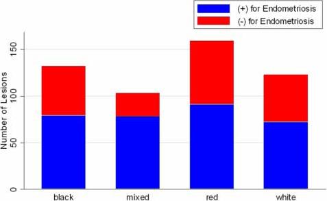
Result(s): Three hundred fifty-seven of 544 lesions believed to be endometriosis by the surgeon had positive histology. Mixed-color lesions most commonly contained endometriosis (76%), with the percentage of positive lesions being similar between single-color groups. Among subtle (red or white) lesions, 58% (164/283) were positive for endometriosis. Thirty women had only red or white lesions, and 18 (60%) had at least one lesion positive for endometriosis. Lesions were most commonly located in the cul-de-sac (64%), utero-sacral ligaments (68%), and ovarian fossa (70%).
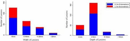
Conclusion(s): Wide, deep, mixed-color lesions in the cul-de-sac, the ovarian fossa, or the utero-sacral ligaments had the highest frequency of endometriosis. More than half of subtle lesions had endometriosis. These results should be considered when diagnosing endometriosis.
Figures
References
-
- Kennedy S, Bergqvist A, Chapron C, D'Hooghe T, Dunselman G, Greb R, et al. ESHRE guideline for the diagnosis and treatment of endometriosis. Hum Reprod. 2005;20:2698–704. - PubMed
-
- Stratton P, Winkel CA, Sinaii N, Merino MJ, Zimmer C, Nieman LK. Location, color, size, depth, and volume may predict endometriosis in lesions resected at surgery. Fertil Steril. 2002;78:743–9. - PubMed
-
- Nisolle M, Donnez J. Peritoneal endometriosis, ovarian endometriosis, and adenomyotic nodules of the rectovaginal septum are three different entities. Fertil Steril. 1997;68:585–96. - PubMed
-
- Martin DC. Endometriosis: correlation between histologic and visual findings at laparoscopy. Am J Obstet Gynecol. 2003;188:1663. author reply −4. - PubMed
-
- Walter AJ, Hentz JG, Magtibay PM, Cornella JL, Magrina JF. Endometriosis: correlation between histologic and visual findings at laparoscopy. Am J Obstet Gynecol. 2001;184:1407–11. discussion 11−3. - PubMed
Publication types
MeSH terms
Substances
Grants and funding
LinkOut - more resources
Full Text Sources
Other Literature Sources
Medical
