. 2007 Sep 5:1167:129-39.
doi: 10.1016/j.brainres.2007.06.046. Epub 2007 Jul 10.

Cytotoxicity of paraquat in microglial cells: Involvement of PKCdelta- and ERK1/2-dependent NADPH oxidase

Affiliations

Cytotoxicity of paraquat in microglial cells: Involvement of PKCdelta- and ERK1/2-dependent NADPH oxidase

Rebecca L Miller et al. Brain Res. .

Abstract

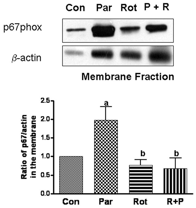
Excess production of reactive oxygen species (ROS) is an important mechanism underlying the pathogenesis of a number of neurodegenerative diseases including Parkinson's disease (PD) which is characterized by a progressive loss of dopaminergic neurons in the substantia nigra. Exposure to paraquat, an herbicide with structure similar to the dopaminergic neurotoxin, 1-methyl-4-phenylpyridinium (MPP+), has been shown to produce PD-like symptoms. Despite previous focus on the dopaminergic neurons and signaling pathways involved in their cell death, recent studies have implicated microglial cells as a major producer of ROS for damaging neighboring neurons. In this study, we examined the source of ROS and the underlying signaling pathway for paraquat-induced cytotoxicity to BV-2 microglial cells. Paraquat-induced ROS production (including superoxide anions) in BV-2 cells was accompanied by translocation of the p67phox cytosolic subunit of NADPH oxidase to the membrane. Paraquat-induced ROS production was inhibited by NADPH oxidase inhibitors, apocynin and diphenylene iodonium (DPI), but not the xanthine/xanthine oxidase inhibitor, allopurinol. Apocynin and DPI also rescued cells from paraquat-induced toxicity. The inhibitors for protein kinase C delta (PKCdelta) or extracellular signal-regulated kinases (ERK1/2) could partially attenuate paraquat-induced ROS production and cell death. Rottlerin, a selective PKCdelta inhibitor, also inhibited paraquat-induced translocation of p67phox. Taken together, this study demonstrates the involvement of ROS from NADPH oxidase in mediating paraquat cytotoxicity in BV-2 microglial cells and this process is mediated through PKCdelta- and ERK-dependent pathways.


Figures

Fig. 1
Fig. 1
Paraquat induced cytotoxicity and increased ROS production in BV-2 microglia cells. A: Paraquat induced a time and dose-dependent increase in ROS production as determined by DCF-DA assay. B: Paraquat induced a time and dose-dependent decrease in cell viability as determined by MTT reduction assay. See Methods for detail description of the protocols. Data are normalized to control and results are mean ± SD from 3 individual experiments.
Fig. 2
Fig. 2
Paraquat caused the increase in superoxide anion as determined by the DHE assay protocol as described in Method. A. Cells were treated with paraquat and/or DDC, a SOD inhibitor, at 37°C for 4 h. Results represent typical fluorescent micrographs depicting BV-2 microglial cells exposed to A-1: control; 2: paraquat (50 μM); 3: DDC (10 μM), and 4: DDC and paraquat. B. Quantitative measurements of paraquat-induced ROS production by DHE in the presence and absence of DDC. Results are mean ± SD from three individual experiments. One-way ANOVA followed by Newman-Keul post-tests indicated significant difference (p < 0.05): a comparing treatment with other groups.
Fig 3
Fig 3
Western blot analysis of p67phox subunit of NADPH oxidase in (A) membrane and (B) cytosol fractions. Microglial cells were exposed to paraquat (50 μM) for 0, 5 and 10 min after which they were subjected to fractionation to separate cytosol and membrane fractions as described in text. Beta-actin was used as loading controls (n=3). Results are representative blots from three independent experiments.
Fig. 4
Fig. 4
Effects of apocynin and DPI on paraquat-induced ROS production and cytotoxity. Microglial cells were treated with paraquat (50 μM) and/or apocynin (1 mM) for (A) ROS determination by DCF at 24 h (n = 6) and (B) assessment of cytotoxicity by MTT at 48 h (n = 3). (C) Treatment of microglial cells with paraquat (50 μM) and/or DPI (1 μM) for 24 h for ROS determination by DCF (n = 5). Results are mean ± SD from number of experiments indicated. One-way ANOVA followed by Newman-Keul post-tests indicated significant difference, p< 0.05: a comparing with control and b comparing with paraquat.
Fig. 5
Fig. 5
The role of PKC on paraquat-induced ROS production and cytotoxicity. Microglial cells were treated with paraquat (50 μM), GF109203x (5 μM), and rottlerin (1 μM) for 24 or 48 h as described above. (A) Treatment with paraquat and/or GF109203x for 24 h for ROS determination; (B) treatment with paraquat and/or GF109203x for 48 h for assessment of cytotoxicity; (C) treatment with paraquat and/or rottlerin for 24 h for ROS determination; and (D) treatment with paraquat and/or rottlerin for 48 h for cytotoxicity. Results are mean ± SD from 3–5 independent experiments. One-way ANOVA followed by Newman-Keul post-tests indicated significant difference, p< 0.05: a comparing with control and b comparing with paraquat.
Fig. 6
Fig. 6
Effects of rottlerin on paraquat-induced translocation of p67 subunit to membranes. Microglial cells were exposed to paraquat and/or rottlerin for 24 h prior to separation of cytosol and membrane fractions for Western blot analysis. Blots were used for densitometer scanning and ratios of p67phox to β-actin in controls were normalized to 1. Results are expressed as mean ± SD (n = 5). One-way ANOVA followed by Newman-Keul post-tests indicated significant difference, p< 0.05: a comparing with control and b comparing with paraquat.
Fig. 7
Fig. 7
Paraquat induces phosphorylation of ERK1/2 and effects of MEK inhibitor on paraquat-induced ROS production and cytotoxicity. (A) For demonstration of ERK1/2 activation, microglial cells were exposed to paraquat (50 μM) for 0, 5 and 10 min prior to lysis for Western blot using antibodies for p-ERK1/2 and total ERK1/2 (n = 3). (B) Cells were exposed to paraquat (50 μM) and/or U0126 (10 μM) for 24 h for assessment of ROS (n = 5); (C) cells were exposed to paraquat (50 μM) and/or U0126 (10 μM) for 48 h for assessment of cytotoxicity (n = 3). One-way ANOVA followed by Newman-Keul post-tests indicated significant difference, p< 0.05: a comparing with control and b comparing with paraquat.
Fig. 8
Fig. 8
A scheme depicting the signaling pathways for paraquat-induced ROS production through activation of NADPH oxidase in microglial cells.

Similar articles

Cited by

References

    1. Abramov AY, Canevari L, Duchen MR. Beta-amyloid peptides induce mitochondrial dysfunction and oxidative stress in astrocytes and death of neurons through activation of NADPH oxidase. Journal of Neuroscience. 2004;24:565–75. - PMC - PubMed
    1. Andresen JJ, Faraci FM, Heistad DD. Vasomotor responses in MnSOD-deficient mice. American Journal of Physiology - Heart & Circulatory Physiology. 2004;287(3):H1141–8. - PubMed
    1. Barcia C, Bahillo AS, Fernandez-Villalba E, Bautista V, Poza MP, Fernandez-Barreiro A, Hirsch EC, Herrero M-T. Evidence of the active microglia in substantia nigra pars compacta of Parkinsonian monkeys 1 year after MPTP exposure. GLIA. 2004;46:402–9. - PubMed
    1. Beal MF. Mitochondria, oxidative damage, and inflammation in Parkinson’s disease. Annals of New York Academy of Sciences. 2003;991:120–31. - PubMed
    1. Bey EA, Xu B, Bhattacharjee A, Oldfield CM, Zhao X, Li Q, Subbulakshmi V, Feldman GM, Wientjes FB, Cathcart MK. Protein kinase C delta is required for p47phox phosphorylation and translocation in activated human monocytes. Journal of Immunology. 2004;173:5730–8. - PubMed

MeSH terms