. 2007 Jul 30:6:7.
doi: 10.1186/1476-5926-6-7.

Evolving concepts of liver fibrogenesis provide new diagnostic and therapeutic options

Affiliations

Evolving concepts of liver fibrogenesis provide new diagnostic and therapeutic options

Olav A Gressner et al. Comp Hepatol. .

Abstract

Despite intensive studies, the clinical opportunities for patients with fibrosing liver diseases have not improved. This will be changed by increasing knowledge of new pathogenetic mechanisms, which complement the "canonical principle" of fibrogenesis. The latter is based on the activation of hepatic stellate cells and their transdifferentiation to myofibroblasts induced by hepatocellular injury and consecutive inflammatory mediators such as TGF-beta. Stellate cells express a broad spectrum of matrix components. New mechanisms indicate that the heterogeneous pool of (myo-)fibroblasts can be supplemented by epithelial-mesenchymal transition (EMT) from cholangiocytes and potentially also from hepatocytes to fibroblasts, by influx of bone marrow-derived fibrocytes in the damaged liver tissue and by differentiation of a subgroup of monocytes to fibroblasts after homing in the damaged tissue. These processes are regulated by the cytokines TGF-beta and BMP-7, chemokines, colony-stimulating factors, metalloproteinases and numerous trapping proteins. They offer innovative diagnostic and therapeutic options. As an example, modulation of TGF-beta/BMP-7 ratio changes the rate of EMT, and so the simultaneous determination of these parameters and of connective tissue growth factor (CTGF) in serum might provide information on fibrogenic activity. The extension of pathogenetic concepts of fibrosis will provide new therapeutic possibilities of interference with the fibrogenic mechanism in liver and other organs.


Figures

Figure 1
Figure 1
Matrix elements and fibrotic changes. Major components of the extracellular matrix (connective tissue) of the liver and the four most important changes in the fibrotic matrix.
Figure 2
Figure 2
Formal pathogenesis of liver fibrosis (fibrogenesis). The "canonical principle" of fibrogenesis starts with necrosis or apoptosis of hepatocytes and inflammation-connected activation of hepatic stellate cells (HSC triggering), their transdifferentiation to myofibroblasts with enhanced expression and secretion of extracellular matrix and matrix deposition (fibrosis). The latter is a precondition for cirrhosis. New pathogenetic mechanisms concern the influx of bone marrow-derived cells (fibrocytes) and of circulating monocytes and their TGF-β driven differentiation to fibroblasts in the damaged liver tissue. A further new mechanism is epithelial-mesenchymal transition (EMT) of bile duct epithelial cells and potentially of hepatocytes. All three complementary mechanisms enlarge the pool of matrix-synthesizing (myo-)fibroblasts in the damaged liver. The most important fibrogenic mediators are transforming growth factor (TGF)-β, platelet-derived growth factor (PDGF), insulin-like growth factor 1 (IGF-1), endothelin-1 (ET-1), and reactive oxygen species (ROS including hydroxyl radicals, superoxid anions). Abbreviations: ASH – alcoholic steatohepatitis; NAFLD – non-alcoholic fatty liver disease. Inset shows an electron micrograph of HSC with numerous lipid droplets indenting the nucleus.
Figure 3
Figure 3
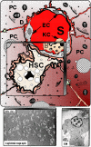
Schematic presentation of hepatic stellate cells (HSC) located in the vicinity of adjacent hepatocytes (PC) beneath the sinusoidal endothelial cells (EC). S – liver sinusoids; KC – Kupffer cells. Down left shows cultured HSC at light-microscopy, whereas at down right electron microscopy (EM) illustrates numerous fat vacuoles (L) in a HSC, in which retinoids are stored.
Figure 4
Figure 4
Compilation of the most important components of extracellular matrix and of mediators synthesized by activated hepatic stellate cells (HSC). Abbreviations: CF – colony-stimulating factor; ET – endothelin; HGF – hepatocyte growth factor; IGF – insulin-like growth factor; KGF – keratinocyte growth factor; LTBP – latent TGF-β binding protein; MCP – monocyte chemotactic peptide; MIP – macrophage inflammatory protein; PAF – platelet activating factor; PDGF – platelet-derived growth factor; PGF – prostaglandin F; SF – scatter factor; TGF – transforming growth factor.
Figure 5
Figure 5
Cellular interactions. Synopsis of cellular interactions of resident liver cells (red) and immigrated inflammatory cells (green) with hepatic stellate cells in the process of activation and transdifferentiation to myofibroblasts. The most important paracrine mediators are given.
Figure 6
Figure 6
Extracellular matrix and TGF-β. Schematic presentation of intracellular TGF-β synthesis, secretion and extracellular immobilization via transglutaminase-dependent fixation of the large latent TGF-β binding protein (LTBP) to extracellular matrix, release by proteases and activation of the latent TGF-β complex by reactive oxygen species (ROS), specific integrins, thrombospondin-1 (TSP-1) or proteases with release of the active TGF-β homodimer, which binds to TGF-β receptors (TβR) III, II, and I to initiate the intracellular signalling cascade of Smad phosphorylation. Regulation of TGF-β occurs at the transcriptional level and, most importantly, by extracellular activation. LAP – latency associated peptide.
Figure 7
Figure 7
Up-to-date mechanisms of fibrogenesis. HSC activation, EMT, influx of fibrocytes, and differentiation of peripheral monocytes to fibroblasts at sites of injury. (Explanation is in the text).

References

    1. Schuppan D, Gressner AM. Function and metabolism of collagens and other extracellular matrix proteins. In: Bircher J, Benhamou JP, McIntyre N, Rizzetto M and Rodés J, editor. Oxford Textbook of Clinical Hepatology. 2. Vol. 2.15. Oxford, Oxford Medical Publications; 1999. pp. 381–407.
    1. Schuppan D, Ruehl M, Somasundaram R, Hahn EG. Matrix as a modulator of hepatic fibrogenesis. Semin Liver Dis. 2001;21:351–72. doi: 10.1055/s-2001-17556. - DOI - PubMed
    1. Gressner AM, Weiskirchen R. Modern pathogenetic concepts of liver fibrosis suggest stellate cells and TGF-beta as major players and therapeutic targets. J Cell Mol Med. 2006;10:76–99. doi: 10.1111/j.1582-4934.2006.tb00292.x. - DOI - PMC - PubMed
    1. de Caestecker MP, Piek E, Roberts AB. Role of transforming growth factor-beta signaling in cancer. J Natl Cancer Inst. 2000;92:1388–1402. doi: 10.1093/jnci/92.17.1388. - DOI - PubMed
    1. Bataller R, Brenner DA. Liver fibrosis. J Clin Invest. 2005;115:209–18. doi: 10.1172/JCI200524282. - DOI - PMC - PubMed

LinkOut - more resources