. 2007 Sep 21;282(38):28149-56.
doi: 10.1074/jbc.M703409200. Epub 2007 Jul 30.

Small heat shock protein alphaA-crystallin regulates epithelial sodium channel expression

Affiliations

Small heat shock protein alphaA-crystallin regulates epithelial sodium channel expression

Ossama B Kashlan et al. J Biol Chem. .

Abstract

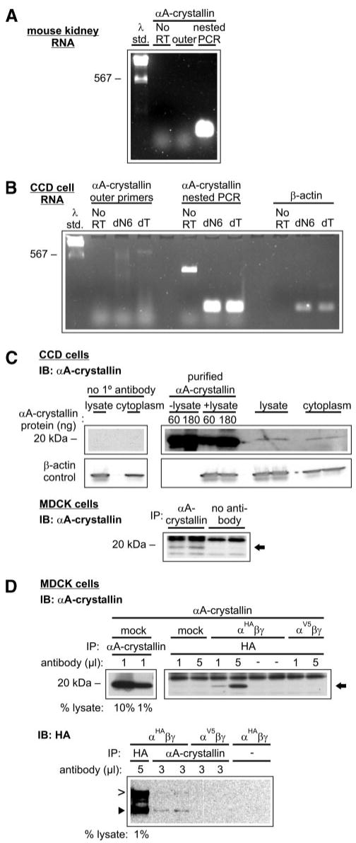
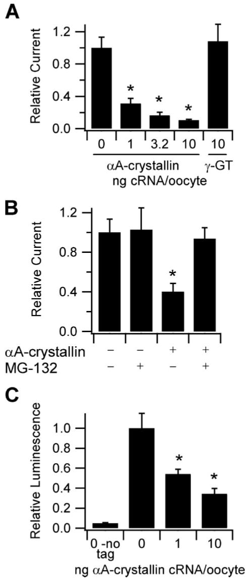
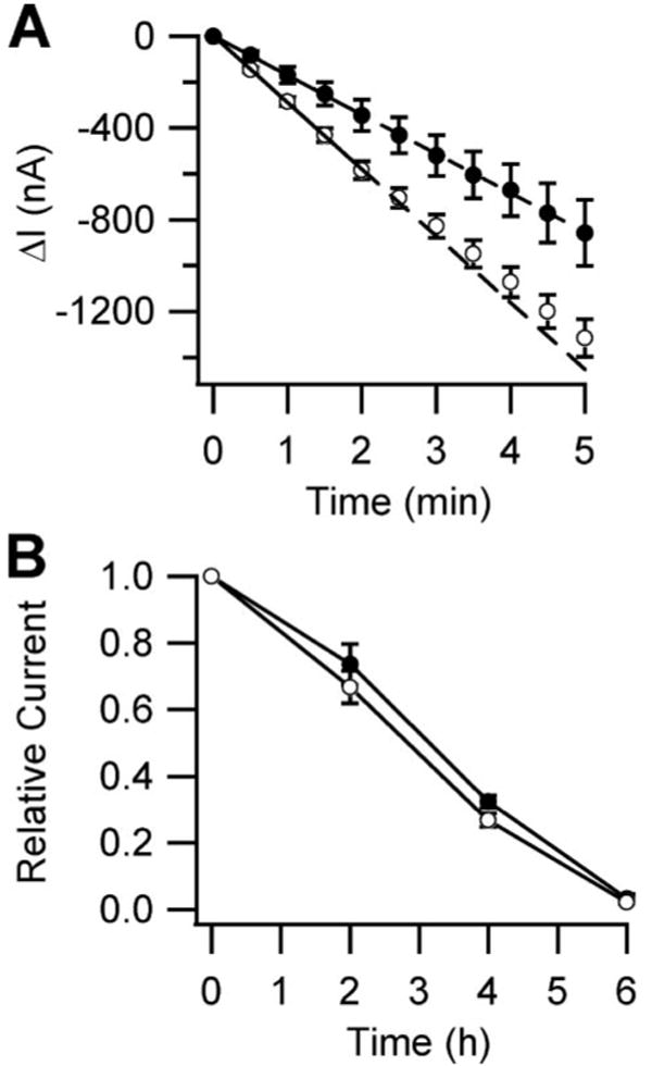
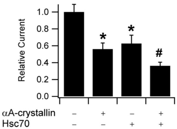
Integral membrane proteins are synthesized on the cytoplasmic face of the endoplasmic reticulum (ER). After being translocated or inserted into the ER, they fold and undergo post-translational modifications. Within the ER, proteins are also subjected to quality control checkpoints, during which misfolded proteins may be degraded by proteasomes via a process known as ER-associated degradation. Molecular chaperones, including the small heat shock protein alphaA-crystallin, have recently been shown to play a role in this process. We have now found that alphaA-crystallin is expressed in cultured mouse collecting duct cells, where apical Na(+) transport is mediated by epithelial Na(+) channels (ENaC). ENaC-mediated Na(+) currents in Xenopus oocytes were reduced by co-expression of alphaA-crystallin. This reduction in ENaC activity reflected a decrease in the number of channels expressed at the cell surface. Furthermore, we observed that the rate of ENaC delivery to the cell surface of Xenopus oocytes was significantly reduced by co-expression of alphaA-crystallin, whereas the rate of channel retrieval remained unchanged. We also observed that alphaA-crystallin and ENaC co-immunoprecipitate. These data are consistent with the hypothesis that small heat shock proteins recognize ENaC subunits at ER quality control checkpoints and can target ENaC subunits for ER-associated degradation.


Figures

FIGURE 1
FIGURE 1. ERAD of ENaC α subunit in yeast requires sHsps for maximal efficiency
ENaC HAαV5 degradation was measured by lysing cells at various times during a chase following cycloheximide addition, and protein levels were quantified by Western analysis and normalized to Sec61 levels. ENaC α subunit degradation was measured for both wild type (○) and mutant (●) yeast, as indicated. n = 6 (hsp26Δhsp42Δ) or 4 (ufd1-1, cim3-1). *, p < 0.05 versus wild type yeast, determined by Student's t test.
FIGURE 2
FIGURE 2. αA-crystallin is expressed in CCD cells and co-immunoprecipitates with ENaC α subunit
αA-crystallin is expressed in mouse kidney tissue and cultured CCD cells (A and B). One microgram of total RNA isolated from native mouse kidney tissue (A) or cultured CCD cells (B) was reverse transcribed using oligo dT (dT) or random hexamers (dN6, CCD cells only) as primers (n = 2). Negative controls were performed in reactions lacking reverse transcriptase (No RT). Predicted reverse transcription-PCR product sizes were 224 bp for αA-crystallin outer primers, 175 bp for αA-crystallin nested primers, and 174 bp for β-actin primers. Note that only nested primers yielded a strong signal of the expected size. C, cultured CCD cell lysates and cytoplasmic extracts were immunoblotted (IB) with mouse anti-αA-crystallin (n = 2). Purified bovine αA-crystallin protein was added as indicated. MDCK cell lysates were immunoprecipitated and immunoblotted with anti-αA-crystallin (n = 3). αA-crystallin is indicated with an arrow. D, ENaC α subunit and αA-crystallin co-immunoprecipitate. MDCK cell extracts were transfected with vectors engineered for the expression of αA-crystallin, ENaC αHA or αV5 subunit, and ENaC β and γ subunits as indicated. Extracts were immunoprecipitated with the indicated amounts of either anti-HA antibody or anti-αA-crystallin antibody. IB: αA-crystallin, n = 4; IB: HA, n = 2. αA-crystallin is indicated with an arrow. The furin-cleaved (65 kDa, ►) and uncleaved (95 kDa, >) ENaC α subunit products are also indicated.
FIGURE 3
FIGURE 3. Effect of αA-crystallin co-expression on the functional expression of ENaC in Xenopus oocytes
A, TEV measurements were performed at −100 mV with oocytes injected with 1 ng each of cRNA encoding the α, β, and γ ENaC subunits and the indicated amounts of cRNA encoding either αA-crystallin or γ-glutamyl transpeptidase (γ-GT). Base-line current was −2.5 μA (n ≥ 16). *, p < 0.0001 versus 0 αA-crystallin, determined by ANOVA. B, effect of proteasomal inhibition on the reduction of ENaC-mediated currents by αA-crystallin. The oocytes were injected with 1 ng of cRNAs encoding each of α, β, and γ ENaC subunits and either water or 6 ng of αA-crystallin cRNA, as indicated. 3 h prior to TEV measurements at −100 mV, half of the oocytes in each group were incubated with 6 μm MG-132. Base-line current was −5.5 μA (n = 15). *, p < 0.05 versus all other groups, determined by ANOVA. C, surface expression of oocytes co-expressing various amounts of αA-crystallin along with wild type α, γ, and βF subunits was determined using anti-FLAG antibodies and a chemiluminescence assay. The oocytes expressing wild type α, β, and γ subunits (no tag) were used to measure background (n ≥ 20 for all groups). *, p < 0.001 versus 0 αA-crystallin, determined by ANOVA.
FIGURE 4
FIGURE 4. Effect of αA-crystallin co-expression on the rates of ENaC cell surface delivery and retrieval in Xenopus oocytes
A, surface delivery rates were determined from oocytes injected with cRNA encoding αS583C, wild type β and γ ENaC subunits, and either water (○) or 6 ng of αA-crystallin cRNA (●). 24 h after injection, the oocytes were treated with MTSET for 4 min, and the currents were measured by TEV at −100 mV every 30 s for 5 min (n = 10). The initial rates were determined by linear regression from the first 2 min for each oocyte. The rates were −290 ± 19 and −170 ± 33 nA/min in the absence and presence of αA-crystallin, respectively (p < 0.05, determined by Student's t test). Wild type base-line current was −7.3 μA. B, surface retrieval rates were determined from oocytes injected with cRNA encoding wild type α, β, and γ ENaC subunits and either water (○) or 6 ng of αA-crystallin cRNA (●). The oocytes were incubated in bath solution alone (data not shown) or bath solution supplemented with 5 μm brefeldin A. The currents were measured every 2 h by TEV at −100 mV (n = 8). The wild type base-line current was −3.3 μA.
FIGURE 5
FIGURE 5. Effect of Hsc70 on the inhibition of ENaC-mediated current by αA-crystallin
TEV measurements were performed at −100 mV. All of the oocytes were injected with 0.5 ng of cRNAs encoding each of α, β, and γ ENaC subunits. 2 ng of cRNA for αA-crystallin and 10 ng of cRNA for Hsc70 were injected as indicated. n = 20 for each experiment. The base-line current was −2.5 μA. *, p < 0.01 versus ENaC alone. #, p < 0.0001 versus ENaC alone, p < 0.05 versus ENaC + Hsc70. p values were determined by ANOVA.

Similar articles

Cited by

References

    1. Garty H, Palmer LG. Physiol Rev. 1997;77:359–396. - PubMed
    1. Rossier BC, Pradervand S, Schild L, Hummler E. Annu Rev Physiol. 2002;64:877–897. - PubMed
    1. Mall M, Grubb BR, Harkema JR, O'Neal WK, Boucher RC. Nat Med. 2004;10:487–493. - PubMed
    1. Kosari F, Sheng S, Li J, Mak DO, Foskett JK, Kleyman TR. J Biol Chem. 1998;273:13469–13474. - PubMed
    1. Firsov D, Gautschi I, Merillat AM, Rossier BC, Schild L. EMBO J. 1998;17:344–352. - PMC - PubMed

Publication types

LinkOut - more resources