. 2007 Aug 6;204(8):1901-9.
doi: 10.1084/jem.20070494. Epub 2007 Jul 30.

The neutrophil serine protease inhibitor serpinb1 preserves lung defense functions in Pseudomonas aeruginosa infection

Affiliations

The neutrophil serine protease inhibitor serpinb1 preserves lung defense functions in Pseudomonas aeruginosa infection

Charaf Benarafa et al. J Exp Med. .

Abstract

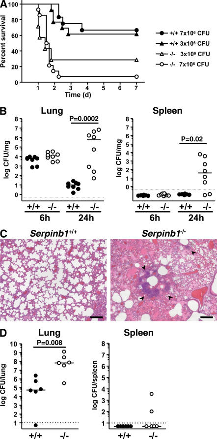
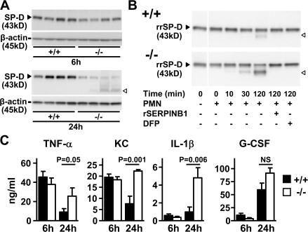
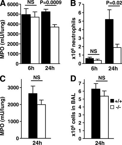
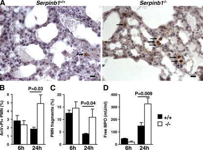
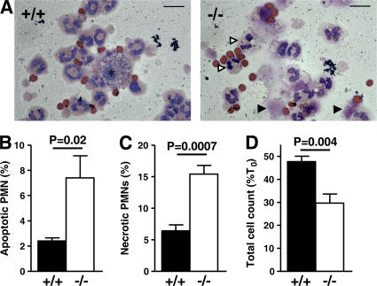
Neutrophil serine proteases (NSPs; elastase, cathepsin G, and proteinase-3) directly kill invading microbes. However, excess NSPs in the lungs play a central role in the pathology of inflammatory pulmonary disease. We show that serpinb1, an efficient inhibitor of the three NSPs, preserves cell and molecular components responsible for host defense against Pseudomonas aeruginosa. On infection, wild-type (WT) and serpinb1-deficient mice mount similar early responses, including robust production of cytokines and chemokines, recruitment of neutrophils, and initial containment of bacteria. However, serpinb1(-/-) mice have considerably increased mortality relative to WT mice in association with late-onset failed bacterial clearance. We found that serpinb1-deficient neutrophils recruited to the lungs have an intrinsic defect in survival accompanied by release of neutrophil protease activity, sustained inflammatory cytokine production, and proteolysis of the collectin surfactant protein-D (SP-D). Coadministration of recombinant SERPINB1 with the P. aeruginosa inoculum normalized bacterial clearance in serpinb1(-/-) mice. Thus, regulation of pulmonary innate immunity by serpinb1 is nonredundant and is required to protect two key components, the neutrophil and SP-D, from NSP damage during the host response to infection.


Figures

Figure 1.
Figure 1.
Generation of serpinb1−/− mice. (A) The WT serpinb1 gene locus comprises seven exons (black rectangles). The targeting plasmid featured a negative selection cassette (DT-A) and a positive/negative selection cassette (Hyg-TK) flanked by two loxP sites (black triangles). The long homology arm also included a third loxP site in intron 6. Targeted ES clones selected for hygromycin resistance but not expressing DT-A were selected for homologous recombination by Southern blotting using an external probe in intron 3 (gray rectangle) after Bgl I restriction. Selected clones were further tested by PCR for the presence of the third loxP site in intron 6 (P1 arrow). ES clones with the deleted locus were identified by PCR (P2 arrow) after transient Cre expression and gancyclovir selection of targeted ES clones. (B) Southern blot of ES clones with WT or targeted loci. (C) PCR P1 was used to select homo logous recombination ES clones that contained the loxP site in intron 6 (denoted as 3 loxP). (D) PCR P2 of mouse tail genomic DNA was used to genotype WT (+/+), heterozygous (+/−), and homozygous (−/−) deleted serpinb1 mice. (E and F) Analysis of bone-marrow neutrophils. (E) Western blot analysis shows the native 42-kD serpinb1 (arrowhead) and the 66-kD serpinb1 complexed with neutrophil proteases (cpx). Protein molecular mass markers are indicated. Lower molecular mass nonspecific bands are also visible. (F) Western blot analysis show 29-kD NE (top) and CG (bottom). Blots were restained for β-actin to indicate equal protein loading.
Figure 2.
Figure 2.
Serpinb1−/− mice fail to clear P. aeruginosa lung infection. (A) Kaplan-Meier survival curves of WT (+/+) and serpinb1-deficient (−/−) mice intranasally inoculated with nonmucoid P. aeruginosa strain PAO1. Increased mortality of serpinb1−/− mice was statistically significant (P = 0.03 at 3 × 10⁶ CFU/mouse; P < 0.0001 at 7 × 10⁶ CFU/mouse). (B) CFUs per milligram of lung (left) and splenic (right) tissue determined 6 and 24 h after inoculation with 3 × 10⁶ CFU P. aeruginosa PAO1 in WT (+/+, filled circles) and serpinb1−/− (−/−, open circles) mice. Each symbol represents a value for an individual mouse. Differences between median values (horizontal lines) were analyzed by the Mann-Whitney U test. Data below the limit of detection (dotted line) are plotted as 0.5 CFU × dilution factor. (C) Lung sections stained with hematoxylin and eosin show bacterial colonies (arrowheads) and alveolar exudate in lungs of serpinb1−/− mice 24 h after infection with P. aeruginosa PAO1. Bars, 50 μm. (D) Total CFUs in the lung and spleen 24 h after inoculation with 2 × 10⁸ CFU of the mucoid P. aeruginosa strain PA M57-15 in WT (+/+, filled circles) and serpinb1−/− (−/−, open circles) mice. Differences between median values (horizontal lines) were analyzed by the Mann-Whitney U test.
Figure 3.
Figure 3.
Proteolysis of SP-D and cytokine production in lungs of P. aeruginosa PAO1–infected mice. (A) Western blot analysis of SP-D (43 kD, black arrowheads) in disulfide-reduced lung homogenate samples at 6 and 24 h after infection. White arrowheads show the SP-D proteolytic cleavage product. Blots were restained for β-actin. (B) Increased in vitro proteolysis of rrSP-D by NSPs of serpinb1−/− PMNs compared with WT PMNs. (C) TNF-α, KC, IL-1β, and G-CSF were measured in BAL fluid of WT (+/+) and serpinb1-deficient (−/−) mice at 6 and 24 h after infection with P. aeruginosa (6 × 10⁶ CFU/mouse; n = 6 per group). Data represent means ± SEM. Representative data from two or more independent experiments are shown (A–C). White lines indicate that intervening lanes have been spliced out.
Figure 4.
Figure 4.
Neutrophil recruitment to the lungs upon challenge with P. aeruginosa and LPS. (A) Neutrophil (PMN) sequestration in lungs assessed by MPO activity in total-lung homogenate and (B) PMN counts in BAL were determined at 6 and 24 h after infection with P. aeruginosa PAO1 (n = 6–8). (C) PMN sequestration and (D) BAL PMN 24 h after intranasal instillation of 10 μg LPS (n = 4–9). Means ± SEM are shown, and data were analyzed by the unpaired t test.
Figure 5.
Figure 5.
Increased death of recruited lung serpinb1−/− neutrophils in vivo. (A) Active caspase-3 staining of leukocytes (arrows) in alveolar space detected by immunohistochemistry at 24 h after infection with P. aeruginosa PAO1. Bars, 20 μm. (B) The percentage of late apoptotic neutrophils (PMNs; AnV+PI+) in BAL of P. aeruginosa–infected mice. (C) PMN fragments defined in flow cytometry by low FSC within the Gr1+ population (Gr1+FSClow). (D) MPO in cell-free BAL supernatant. Means ± SEM are shown (n = 4–6), and the data were analyzed by the unpaired t test.
Figure 6.
Figure 6.
Increased apoptosis and necrosis of serpinb1−/− neutrophils in vitro. (A) Representative cytospins of LPS-recruited BAL cells after 24 h of in vitro culture. Macrophages are indicated by an asterisk, and apoptotic and lytic PMNs are indicated by white and black arrowheads, respectively. Bars, 10 μm. The percentage of PMNs with characteristic apoptotic (B) or lytic (C) morphologies relative to total PMNs after 24 h of in vitro culture. (D) The percentage of cells remaining after 24 h in culture. The means ± SEM of five experiments are shown, and data were analyzed by the unpaired t test (B–D).

Similar articles

Cited by

References

    1. Reeves, E.P., H. Lu, H.L. Jacobs, C.G. Messina, S. Bolsover, G. Gabella, E.O. Potma, A. Warley, J. Roes, and A.W. Segal. 2002. Killing activity of neutrophils is mediated through activation of proteases by K+ flux. Nature. 416:291–297. - PubMed
    1. Belaaouaj, A., K.S. Kim, and S.D. Shapiro. 2000. Degradation of outer membrane protein A in Escherichia coli killing by neutrophil elastase. Science. 289:1185–1188. - PubMed
    1. Belaaouaj, A., R. McCarthy, M. Baumann, Z. Gao, T.J. Ley, S.N. Abraham, and S.D. Shapiro. 1998. Mice lacking neutrophil elastase reveal impaired host defense against gram negative bacterial sepsis. Nat. Med. 4:615–618. - PubMed
    1. Tkalcevic, J., M. Novelli, M. Phylactides, J.P. Iredale, A.W. Segal, and J. Roes. 2000. Impaired immunity and enhanced resistance to endotoxin in the absence of neutrophil elastase and cathepsin G. Immunity. 12:201–210. - PubMed
    1. Chmiel, J.F., and P.B. Davis. 2003. State of the art: why do the lungs of patients with cystic fibrosis become infected and why can't they clear the infection? Respir. Res. 4:8. - PMC - PubMed

Publication types

MeSH terms