. 2007 Jul 30;178(3):345-54.
doi: 10.1083/jcb.200701163.

Astrin is required for the maintenance of sister chromatid cohesion and centrosome integrity

Affiliations

Astrin is required for the maintenance of sister chromatid cohesion and centrosome integrity

Kerstin H Thein et al. J Cell Biol. .

Abstract

Faithful chromosome segregation in mitosis requires the formation of a bipolar mitotic spindle with stably attached chromosomes. Once all of the chromosomes are aligned, the connection between the sister chromatids is severed by the cysteine protease separase. Separase also promotes centriole disengagement at the end of mitosis. Temporal coordination of these two activities with the rest of the cell cycle is required for the successful completion of mitosis. In this study, we report that depletion of the microtubule and kinetochore protein astrin results in checkpoint-arrested cells with multipolar spindles and separated sister chromatids, which is consistent with untimely separase activation. Supporting this idea, astrin-depleted cells contain active separase, and separase depletion suppresses the premature sister chromatid separation and centriole disengagement in these cells. We suggest that astrin contributes to the regulatory network that controls separase activity.


Figures
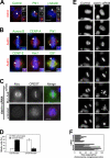
Figure 1.
Figure 1.
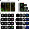
Astrin depletion results in impaired chromosome alignment and the formation of multipolar spindles. (A) Pro- and prometaphase HeLa cells were stained with antibodies against astrin and centrin, Plk1, or γ-tubulin. Centrosomes indicated with arrows are shown enlarged in the insets. (B) Metaphase HeLa cells were processed for immunofluorescence using antibodies against astrin and aurora B, CENP-A, Plk1, CENP-E, Hec1, or EB1. The kinetochores indicated with arrows are shown enlarged in the insets. (C) HeLa cells were transfected with myc-astrin constructs containing five silent mutations in the sequence targeted by the astrin siRNA oligonucleotide 24 h before transfection with astrin siRNA oligonucleotides and stained with CREST antiserum and antibodies against myc. (D) The percentage of transfected and untransfected cells displaying the astrin depletion phenotype was scored in two independent experiments. Error bars represent SD. (E) Stills of representative videos of control and astrin-depleted cells. Time is indicated in minutes. All astrin-depleted cells analyzed (n = 10) exhibited delayed chromosome congression (compare t = 24 min in control and astrin siRNA cells) and unstable metaphase plates. Note the unaligned chromosomes in astrin siRNA at t = 104 min and t = 160 min (arrowheads). The depicted astrin-depleted cell is initially bipolar but forms a multipolar spindle (asterisks indicate spindle poles) during the mitotic arrest. (F) Control or astrin-depleted HeLa S3 cells expressing histone-H2B-GFP were followed by live cell analysis. The time required for chromosome congression was plotted. Bars, 10 μm.
Figure 2.
Figure 2.
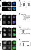
Astrin-depleted cells are spindle checkpoint arrested. (A–D) Control or astrin-depleted HeLa cells were stained with antibodies against α-tubulin (green) and either Mad2 or BubR1 (red; A and B) or were stained with CREST antiserum (red) and antibodies against cyclin B1 or securin (green; C and D), respectively. Mad2- or strongly BubR1-positive kinetochores are indicated by arrowheads. DNA was visualized with DAPI. (E) The levels of securin staining in control or astrin-depleted mitotic cells were measured in four independent experiments using ImageJ software, analyzing 15–20 cells of each kind per experiment. (F) Lysates of mitotic control and astrin-depleted HeLa cells harvested by mitotic shake off were immunoblotted with antibodies against the indicated proteins. The two astrin bands appear as one because of a higher percentage of SDS gel used. (G) HeLa cells were transfected with astrin siRNA and 16 h later with Mad2 or control siRNA oligonucleotides for an additional 24 h, and the mitotic index was scored. (H) Control or astrin-depleted cells were treated with DMSO or 10 μM ZM447493 for 3 h, and the mitotic index was scored. Error bars represent SD. Bars, 10 μm.
Figure 3.
Figure 3.
Microtubule–kinetochore interactions are unstable, and outer kinetochore components are mislocalized in astrin-depleted cells. (A) HeLa control cells and astrin- or Hec1-depleted cells were incubated on ice for 20 min before fixation. Cold-stable K-fiber microtubules were revealed by kinetochore staining with CREST serum and antibodies against α-tubulin. The boxed areas are shown magnified in the bottom panels. Note the presence of unattached kinetochores in astrin-depleted cells. Bars (top), 10 μm; (bottom) 1 μm. (B) Control or astrin-depleted HeLa cells were preextracted before fixation, and microtubule–kinetochore interactions were visualized as in A. (C) Control (left) and astrin-depleted HeLa cells (right) were stained for astrin (red) and either Hec1, Bub1, CENP-E, or CENP-F (green). (B and C) Bars, 10 μm.
Figure 4.
Figure 4.
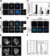
Astrin depletion causes the loss of centriole and sister chromatid cohesion. (A) Control or astrin-depleted cells were stained with antibodies against centrin (red) and α-tubulin (green). DNA was visualized with DAPI. Individual centrosomes are shown enlarged on the right. (B) HeLa cells depleted of aurora B or TOGp were stained as in A. (C) Quantitative analysis of the centriole number at the poles of multipolar spindles in cells depleted of astrin, aurora B, or TOGp. The number of poles containing no, one, or two centrin stainings was plotted as a percentage of the total number of poles. The cells were analyzed double blind by three different researchers. The graphs represent triplicate experiments of at least 40 cells each. (D) Astrin-, aurora B–, and TOGp-depleted cells were stained with antibodies against pericentrin (red) and α-tubulin (green). The arrowheads indicate acentriolar spindle poles in the TOGp-depleted cell. (E) Chromosome spreads were prepared from mitotic HeLa cells depleted of Gl2 (control), CENP-E (both 48-h RNAi), Sgo1, and astrin (both 40-h RNAi). Representative pictures of each sample are shown. (F) Quantitation of the chromosome spreads shown in E. Each bar represents triplicate experiments. 100 cells of each sample were counted per experiment. Error bars represent SD. Bars, 10 μm.
Figure 5.
Figure 5.
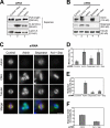
Separase-dependent formation of multipolar spindles and loss of sister chromatid cohesion in cells depleted of astrin. (A) Extracts prepared from nocodazole-arrested HeLa control cells (lane 1), control cells that had been released from a nocodazole block for 100 min (lane 2), or astrin-depleted cells harvested by mitotic shake off (lane 3) were immunoblotted with mouse anti–C-separase antibodies and mouse anti–lamin A as a loading control. The blot shown is a representative example of five independent experiments. (B) HeLa cells were depleted of astrin and separase individually or together. Extracts from these and control cells were blotted for astrin and separase and tubulin as a loading control. Note that astrin-depleted cells (lane 3) contain more separase than asynchronous (lane 1) but similar amounts to nocodazole-arrested control cells (lane 2) because of the elevated mitotic index. (C) Cells treated as in B were stained with antibodies against astrin (red) and α-tubulin (green). Representative images of each cell population are shown. (D–F) Quantitation of the mitotic index, percentage of multipolar cells of mitotic cells, and degree of sister chromatid separation observed in astrin-, separase-, or double-depleted cells and control cells. Error bars represent SD. Bar, 10 μm.

Similar articles

Cited by

References

    1. Baumann, C., R. Korner, K. Hofmann, and E.A. Nigg. 2007. PICH, a centromere-associated SNF2 family ATPase, is regulated by Plk1 and required for the spindle checkpoint. Cell. 128:101–114. - PubMed
    1. Cassimeris, L., and J. Morabito. 2004. TOGp, the human homolog of XMAP215/Dis1, is required for centrosome integrity, spindle pole organization, and bipolar spindle assembly. Mol. Biol. Cell. 15:1580–1590. - PMC - PubMed
    1. Chan, G.K., B.T. Schaar, and T.J. Yen. 1998. Characterization of the kinetochore binding domain of CENP-E reveals interactions with the kinetochore proteins CENP-F and hBUBR1. J. Cell Biol. 143:49–63. - PMC - PubMed
    1. Chang, M.S., C.J. Huang, M.L. Chen, S.T. Chen, C.C. Fan, J.M. Chu, W.C. Lin, and Y.C. Yang. 2001. Cloning and characterization of hMAP126, a new member of mitotic spindle-associated proteins. Biochem. Biophys. Res. Commun. 287:116–121. - PubMed
    1. Ciosk, R., W. Zachariae, C. Michaelis, A. Shevchenko, M. Mann, and K. Nasmyth. 1998. An ESP1/PDS1 complex regulates loss of sister chromatid cohesion at the metaphase to anaphase transition in yeast. Cell. 93:1067–1076. - PubMed

Publication types