. 2007 Jul 30;178(3):465-76.
doi: 10.1083/jcb.200702009.

Divergent functions and distinct localization of the Notch ligands DLL1 and DLL3 in vivo

Affiliations

Divergent functions and distinct localization of the Notch ligands DLL1 and DLL3 in vivo

Insa Geffers et al. J Cell Biol. .

Abstract

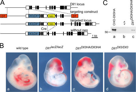
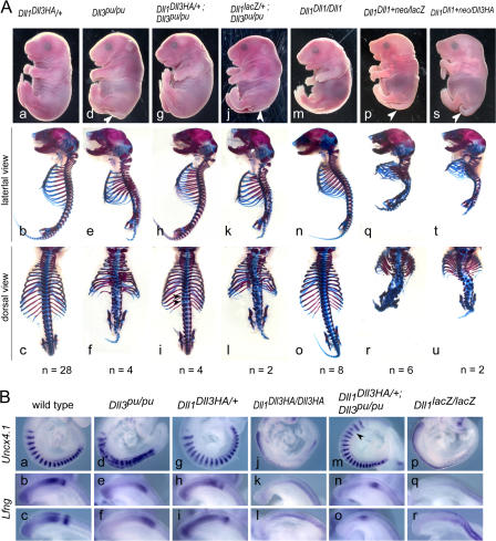
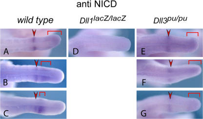
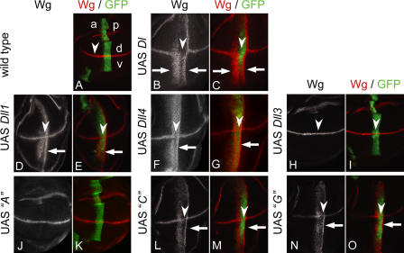
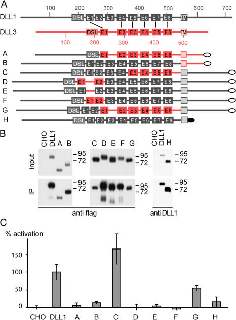
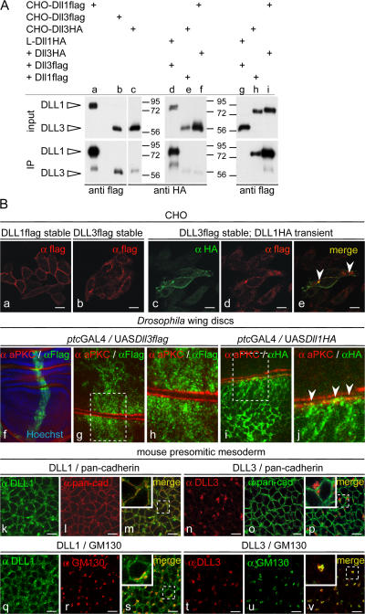
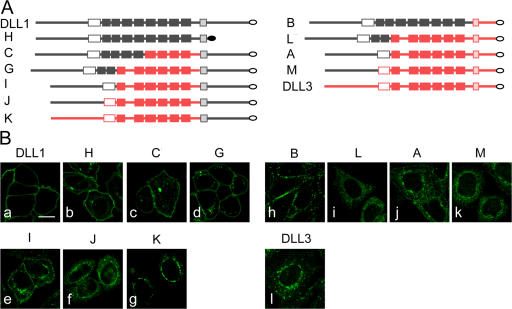
The Notch ligands Dll1 and Dll3 are coexpressed in the presomitic mesoderm of mouse embryos. Despite their coexpression, mutations in Dll1 and Dll3 cause strikingly different defects. To determine if there is any functional equivalence, we replaced Dll1 with Dll3 in mice. Dll3 does not compensate for Dll1; DLL1 activates Notch in Drosophila wing discs, but DLL3 does not. We do not observe evidence for antagonism between DLL1 and DLL3, or repression of Notch activity in mice or Drosophila. In vitro analyses show that differences in various domains of DLL1 and DLL3 individually contribute to their biochemical nonequivalence. In contrast to endogenous DLL1 located on the surface of presomitic mesoderm cells, we find endogenous DLL3 predominantly in the Golgi apparatus. Our data demonstrate distinct in vivo functions for DLL1 and DLL3. They suggest that DLL3 does not antagonize DLL1 in the presomitic mesoderm and warrant further analyses of potential physiological functions of DLL3 in the Golgi network.


Figures

Figure 1.
Figure 1.
Generation of Dll1Dll3Haki, Dll1Dll3ki, and Dll1Dll1ki mice. (A) Targeting strategy for introduction of Dll3 and Dll1 into the Dll1 locus. White and black boxes indicate noncoding and coding regions, respectively. The blue box indicates a Dll1, Dll3, or Dll3HA cDNA, respectively. DT, diphtheria toxin A chain. (B) E10.5 embryos. (C) Verification of Dll3HA expression by Western blot analysis with anti-HA antibodies. Lysates of CHO cells transfected with Dll3HA cDNA (a) and of wild type (b) and Dll1Dll3HA/Dll3HA (c) E9.5 embryos. Embryo lysates represent half of one embryo, respectively.
Figure 2.
Figure 2.
Phenotypes and somite patterning of embryos with various Dll1 and Dll3 allele combinations. (A) Embryo appearance (top) and skeletal preparations (bottom) of E18.5 embryos with the genotypes indicated on top. Dll1 Dll3HA/+ embryos (a–c) have an essentially normal axial skeleton despite an increased gene dosage ratio of Dll3 to Dll1. Skeletal defects of homozygous Dll3-null mutants (d–f) are rescued by Dll3HA expressed from the Dll1 locus (g–i) except for minor residual defects (i, arrowheads). Increased gene dosage ratio of Dll3 to Dll1 does not enhance a hypomorphic Dll1 phenotype (compare p–r with s–u). Note that modification of the Dll1 locus does not lead to any phenotypic alterations in the skeleton (m–o). (B) A-P somite patterning at E9.5 indicated by Uncx4.1 expression and dynamic Lfng expression patterns are indistinguishable in wild-type (a–c) and Dll1 Dll3HA/+ embryos (g–i). Abnormal expression of Uncx4.1 and Lfng in homozygous Dll3pu-null mutants (d–f) is restored by Dll3HA expressed from the Dll1 locus (m–o) except for minor irregularities of Uncx4.1 expression (m, arrowhead). Expression of Dll3HA instead of Dll1 (j–l) does not rescue the patterning defects of Dll1-null mutants (p–r).
Figure 3.
Figure 3.
NICD expression in the PSM of Dll3 mutant embryos. Whole-mount immunohistochemistry for activated Notch 1 (NICD) readily detected oscillating activity in E10.5 embryo wild-type tails (A–C; n = 13). In Dll1-null mutant tails (D), NICD was not detected in the PSM (n = 4), whereas in Dll3-null mutant tails (E–G), the pattern of Notch1 activity is static (n = 14) with no obvious up-regulation of NICD protein levels.
Figure 4.
Figure 4.
Activity of Dll1 and Dll3 in D. melanogaster wing discs. Expression of D. melanogaster Dl and vertebrate orthologues with ptcGal4 in the wing imaginal discs. (A) Expression of Wg and ptcGal4 in a wing imaginal disc of the late third larval instar. Wg (red) is induced along the dorsoventral (d-v) compartment boundary by Notch signaling. Expression of UAS GFP (green) reveals the stripe-like expression domain of ptcGal4, which runs perpendicular to the Wg domain at the anterior side of the A-P compartment border. Expression within the ptcGal4 domain increases toward the posterior (right). (B and C) Ectopic Wg activation along the A-P boundary (arrows) induced by ectopic expression of D. melanogaster Dl. Two ectopic stripes of Wg expression are induced (arrows). The broader, anterior-located stripe is in the region of low Dl expression. The second thinner is induced in cells adjacent to the ptc domain. In the region with highest expression of Dl, expression of Wg is not induced because of the cis-inhibitory effect of Dl at high levels of expression. Note that high levels of Dl also suppress Notch activity at the dorsoventral boundary as indicated by down-regulation of Wg (arrowheads). Expression of Dll1flag (D and E) or Dll4flag (F and G) along the A-P boundary activates ectopic expression of Wg (arrows) in a pattern similar to Dl. However, the cis-inhibitory effect is weaker than in the case of Dl. (H and I) Expression of Dll3flag has no effect on the activity of the Notch pathway in D. melanogaster. In addition, endogenous expression of Wg along the dorsoventral border is not affected (arrowheads). (J and K) Expression of construct A (Dll1/Dll3 chimera; Fig. 5) does not activate the Notch pathway in D. melanogaster. (L and M) Ectopic Wg activation along the A-P wing border (arrow) by ectopic expression of construct C (Dll1/Dll3 chimera; Fig. 5). In the regions with highest expression, construct C slightly suppresses Notch activity, as indicated by the slight down-regulation of Wg in its normal expression domain (arrowheads). (N and O) Ectopic Wg activation along the A-P wing border (arrows) by ectopic expression of construct G (Dll1/Dll3 chimera; Fig. 5). In the regions with highest expression, construct G slightly suppresses Notch activity, as indicated by the slight down-regulation of Wg in its normal expression domain (arrowheads). Note that construct C, as well as construct G, signals to cells adjacent to the posterior domain boundary (L–O, arrows).
Figure 5.
Figure 5.
Analysis of Dll1-Dll3 chimeric ligands. (A) Schematic overview of wild-type DLL1 and DLL3 and chimeric constructs used to generate stably expressing CHO cell lines. DLL1 protein is shown in black and DLL3 in red. Numbers indicate the amino acid residue numbers. DSL, DSL domain; E1–E8, EGF-like repeats; the flag tag is indicated by gray ovals and the HA tag in construct H by a black oval. Corresponding EGF repeats of DLL1 and DLL3 are connected by black lines. (B) Western blot analysis of cell lysates (input) and streptavidin immunoprecipitated protein after surface biotinylation (IP). CHO cells stably expressing chimeric ligands show similar (input A and B) or even more (input C–H) expression compared with DLL1-expressing cells. All chimeric ligands are present on the cell surface (IP), chimeric ligands A, C, and G at lower levels and chimeric ligands B, D–F, and H at similar or even higher levels compared with DLL1. (C) Notch transactivation assays. CHO cells stably expressing DLL1 and chimeric ligands as shown in panel A were cocultivated with Notch1-HeLa cells transfected with the (RbpJ)6-luciferase reporter gene. Luciferase activity (percentage of activation) of chimeric ligands A–H was measured against negative (CHO wild-type cells) and positive (CHO-Dll1 cells) controls set to 0 and 100% relative activation, respectively. Four cocultivations were performed per construct and analyzed in two independent experiments each, including negative and positive controls.
Figure 6.
Figure 6.
Localization of DLL1 and DLL3 proteins. (A) Western blot analysis of cell lysates (input) and streptavidin-immunoprecipitated protein after surface biotinylation (IP). CHO cells stably expressing DLL3 (b and c) at amounts similar to cells expressing DLL1 (a) present significantly less DLL3 on the surface. L cells (mouse fibroblast cell line) coexpressing DLL3flag at significantly higher levels than DLL1HA (compare input lanes d and g) present DLL1 efficiently on the surface but not DLL3 (compare IP lanes d and g). CHO cells coexpressing DLL3HA and DLL1flag (compare input lanes e and f with h and i) present DLL1 efficiently on the surface but DLL3 only in trace amounts (compare IP lanes e and f with h and i). (B) Detection of DLL1 and DLL3 by immunofluorescence. (a–e) Localization of DLL1 and DLL3 in overexpressing CHO cells. CHO cells expressing DLL1 (a and c) show a clear cell surface staining, whereas DLL3 (b and d) is detected almost exclusively inside the cell. DLL1 and DLL3 colocalize only in some vesicular structures (e, arrowheads) but not significantly at the membrane. (f–j) Localization of DLL1 and DLL3 in D. melanogaster wing disc cells. (f) Overview of a wing disc stained for the apical cell membrane marker aPKC and DLL3flag, and Hoechst staining to visualize nuclei. (g and h) Confocal images of two opposed apical cell membranes (red) of an epithelial fold in panel f. DLL3 is found in intracellular granules or vesicles. (i and j) Confocal images of two opposed apical cell membranes (red) of a wing disc stained for aPKC and DLL1flag. DLL1 outlines cell membranes and colocalizes at the apical membrane with aPKC (j, arrowheads). (k–v) Immunofluorescent detection of DLL1 and DLL3 in PSM cells of E9.5 embryos. Endogenous DLL1 is present at the surface (k) and colocalizes with the membrane (m) and in vesicular structures with the cis-Golgi marker GM130 (s). DLL3 does not localize to the membrane (n) and does not colocalize with anti-pancadherin staining (p) but is detected in vesicular structures in the cytoplasm (t), mostly overlapping with GM130 (v). Bars, 10 μm.
Figure 7.
Figure 7.
Localization of chimeric DLL1 and DLL3 proteins. (A) Schematic representation of chimeric proteins containing the ICD of DLL1 (left) or DLL3 (right) arranged according to the extent of extracellular DLL1 sequences (top to bottom). Gray parts and red parts indicate DLL1 and DLL3 sequences, respectively. White filling indicates DSL domains, light gray or red shading indicates TMs, and filled boxes indicate EGF repeats. (B) Confocal images of CHO cells stably expressing chimeric ligands and stained by indirect immunofluorescence. Similar to DLL1 (a) and DLL1 lacking the ICD (b), chimeric ligands that contained the TM-ICD of DLL1 and the DLL1 N-terminal portion including the DSL domain were detected on the cell surface (c–e), in addition to some variable intracellular expression. Presence of the DLL1 N terminus alone was not sufficient to direct detectable surface expression (f), similar to the extracellular domain of DLL3 fused to DLL1 TM-ICD (g). Surface presentation of a chimera containing the DLL1 extracellular domain juxtaposed to the DLL3 TM-ICD (h). Chimeras that contain the DLL1 N-terminal portion including the DSL domain, and the DLL3 TM in the context of juxtaposed DLL3 intra- and extracellular sequences were retained intracellularly (i and j). Intracellular localization of DLL3 with the N terminus replaced by the corresponding DLL1 sequence, and of DLL3, respectively (k and l). A–C, G, and H refer to chimeras shown in Fig. 5 A and I–M to additional ones. Chimera H (b) was detected with anti-DLL1 and all other chimeras with anti-flag antibodies. Bar, 10 μm.

References

    1. Abdelkhalek, H.B., A. Beckers, K. Schuster-Gossler, M.N. Pavlova, H. Burkhardt, H. Lickert, J. Rossant, R. Reinhardt, L.C. Schalkwyk, I. Müller, et al. 2004. The mouse homeobox gene Not is required for caudal notochord development and affected by the truncate mutation. Genes Dev. 18:1725–1736. - PMC - PubMed
    1. Artavanis-Tsakonas, S., M.D. Rand, and R.J. Lake. 1999. Notch signaling: cell fate control and signal integration in development. Science. 284:770–776. - PubMed
    1. Ascano, J.M., L.J. Beverly, and A.J. Capobianco. 2003. The C-terminal PDZ- ligand of JAGGED1 is essential for cellular transformation. J. Biol. Chem. 278:8771–8779. - PubMed
    1. Barsi, J.C., R. Rajendra, J.I. Wu, and K. Artzt. 2005. Mind bomb1 is a ubiquitin ligase essential for mouse embryonic development and Notch signaling. Mech. Dev. 122:1106–1117. - PubMed
    1. Blaumueller, C.M., and S. Artavanis-Tsakonas. 1997. Comparative aspects of Notch signaling in lower and higher eukaryotes. Perspect. Dev. Neurobiol. 4:325–343. - PubMed

Publication types

MeSH terms