. 2007 Jul 30;178(3):503-16.
doi: 10.1083/jcb.200609146.

The neurite outgrowth multiadaptor RhoGAP, NOMA-GAP, regulates neurite extension through SHP2 and Cdc42

Affiliations

The neurite outgrowth multiadaptor RhoGAP, NOMA-GAP, regulates neurite extension through SHP2 and Cdc42

Marta Rosário et al. J Cell Biol. .

Abstract

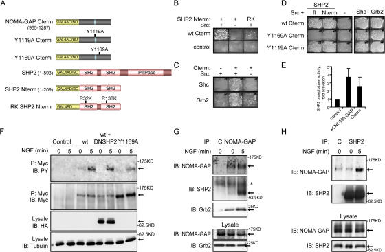
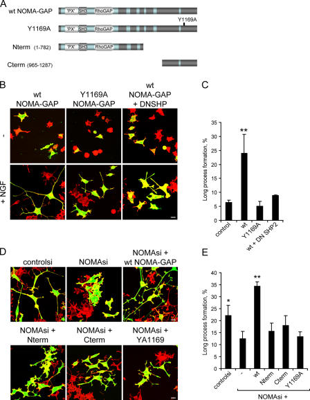
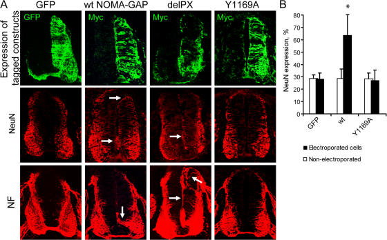
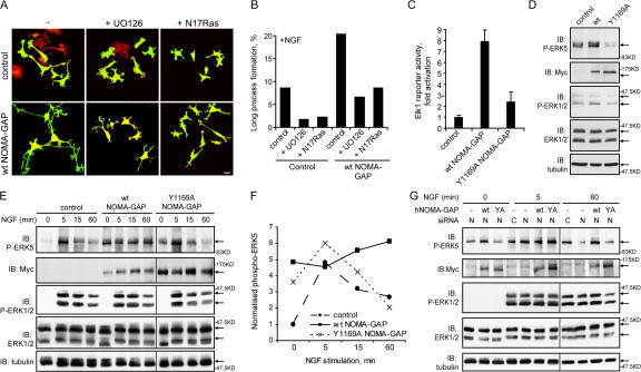
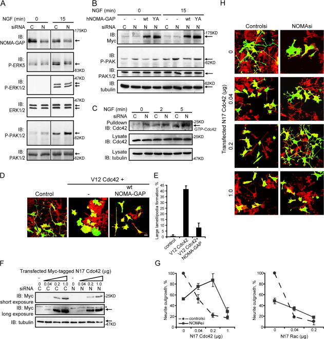
Neuronal differentiation involves the formation and extension of neuronal processes. We have identified a novel regulator of neurite formation and extension, the neurite outgrowth multiadaptor, NOMA-GAP, which belongs to a new family of multiadaptor proteins with RhoGAP activity. We show that NOMA-GAP is essential for NGF-stimulated neuronal differentiation and for the regulation of the ERK5 MAP kinase and the Cdc42 signaling pathways downstream of NGF. NOMA-GAP binds directly to the NGF receptor, TrkA, and becomes tyrosine phosphorylated upon receptor activation, thus enabling recruitment and activation of the tyrosine phosphatase SHP2. Recruitment of SHP2 is required for the stimulation of neuronal process extension and for sustained activation of ERK5 downstream of NOMA-GAP. In addition, we show that NOMA-GAP promotes neurite outgrowth by tempering activation of the Cdc42/PAK signaling pathway in response to NGF. NOMA-GAP, through its dual function as a multiadaptor and RhoGAP protein, thus plays an essential role downstream of NGF in promoting neurite outgrowth and extension.


Figures

Figure 1.
Figure 1.
Structure and expression pattern of NOMA-GAP and RICS. (A) Schematic representation of the structure of human NOMA-GAP and RICS proteins. Phox (PX), Phox-like (‘PX’), SH3, and RhoGAP domains are indicated. Additional homology regions are shaded light blue. The degree of amino acid identity of particular domains between the proteins is given in percentages. Novel RICS N-terminal sequences are indicated by a thick blue line. (B) Expression of NOMA-GAP mRNA (blue-purple) is shown in whole mount and in mid-thoracic transverse sections of murine embryos (E8.5 to E11.5). Multiple probes to distinct portions of the NOMA-GAP and RICS gene were tested, with similar results. Sections from E11.5 embryos have been counterstained with Nuclear Fast Red (pink). Immunostaining for neurofilament (NF) of an adjacent section is shown in red. (C) Localization of endogenous NOMA-GAP protein (green) in transverse sections of mouse embryos (E11). NF staining is shown in red. (D) Localization of endogenous NOMA-GAP in NGF-stimulated PC12 cells. NOMA-GAP staining is shown in green. The two bottom panels show magnifications of the growth cones of PC12 cells stimulated with NGF for 5 d and co-stained for endogenous NOMA-GAP (green) and either total membrane or polymerized actin as indicated (red). Filled arrows indicate selected points of NOMA-GAP accumulation; empty arrows indicate microspikes emanating from neurite tips.
Figure 2.
Figure 2.
NOMA-GAP is required for NGF-stimulated process extension. (A) Down-regulation of endogenous NOMA-GAP protein in NGF-stimulated PC12 cells transfected with control siRNA (controlsi) or siRNA directed against rat NOMA-GAP (NOMAsi). NOMA-GAP immunoprecipitates (IP) were immunoblotted (IB) for NOMA-GAP. Lysates were also immunoblotted for ERK1/2. (B) Down-regulation of immunofluorescent staining for endogenous NOMA-GAP in NGF-stimulated siRNA-transfected PC12 cells. Cells were transfected with control siRNA or NOMA-GAP siRNA in the presence of limiting amounts of the transfection marker, GFP, and immunostained for endogenous NOMA-GAP. Mean pixel intensity of NOMA-GAP staining per GFP-positive cell is shown. The mean staining of controlsi (n = 40) and NOMAsi (n = 56) samples were compared with a t test for unequal variances and found to be significantly different (P < 0.0005; see Materials and methods). (C) Effect of down-regulation of NOMA-GAP expression on NGF-stimulated differentiation in PC12 cells. Cells transfected with control siRNA, NOMA-GAP siRNA, or RICS siRNA were stained for polymerized actin (red) and the transfection marker GFP (green) 72 h after transfection. Arrows indicate selected transfected cells. Bar, 20 μm. (D) Quantification of neuronal process extension in siRNA-transfected PC12 cells (five independent experiments). Total processes, cells bearing processes >30 μm long; long processes, cells bearing processes >100 μm long. The means of the four samples were compared with the multiple test of ANOVA and found to be significantly different (P < 0.0005). The error probabilities of pairwise tests were corrected by Bonferroni. The means of the control and NGF+NOMAsi samples were found to be significantly different from the mean of the NGF+controlsi sample (P < 0.0005 in both cases for Total processes and P < 0.0005 and P < 0.001, respectively for Long processes; summarized by asterisks; see Materials and methods). Percentages denote level of inhibition.
Figure 3.
Figure 3.
NOMA-GAP interacts with TrkA. (A) Coimmunoprecipitation of endogenous NOMA-GAP and TrkA from NGF-stimulated PC12 cells. TrkA immunoprecipitates were immunoblotted (IB) as indicated. (B) Diagrammatic representation of the GAL4 activation domain: NOMA-GAP fusion constructs used for yeast two-hybrid analysis. (C) Interaction of huNOMA-GAP with the cytoplasmic domain of huTrkA in the yeast two-hybrid system. The growth of transformed yeast on plates selecting for protein interaction (Selection plate) or for the presence of both plasmids (Total growth plate) is shown.
Figure 4.
Figure 4.
NOMA-GAP stimulates the extension of long neuronal processes. (A) Schematic representation of wild-type (wt) and deletion mutant NOMA-GAP constructs. (B) NOMA-GAP stimulates the extension of long neuronal processes in PC12 cells stimulated with low levels of NGF for 48 h. Samples were stained as described in Fig. 2 C. (C) Summary of the average percentage of GFP-positive cells bearing short or long neuronal processes in three independent experiments. (D) Quantification of the effect of different NOMA-GAP deletion mutants on the extension of long neuronal processes in three independent experiments. The means of the different samples were compared as described in Fig. 2 D. The means of the wt and delPX samples were found to be significantly different from the mean of the control sample (P < 0.01 and P < 0.0005, respectively).
Figure 5.
Figure 5.
The C terminus of NOMA-GAP acts as a docking site for multiple signaling effectors. (A) Schematic representation of the NOMA-GAP and SHP2 constructs used for yeast two-hybrid analysis (where C-term refers to C terminus of NOMA-GAP; fl to full-length SHP2; Nterm to the N terminus of SHP2). (B–D) Yeast growth under selection for the reconstitution of GAL4 transcriptional activity. (B) Interaction of NOMA-GAP Cterm with wild-type and RK mutant SHP2 Nterm. (C) Interaction of NOMA-GAP Cterm with Shc and Grb2. (D) Interaction of point mutants of NOMA-GAP Cterm with SHP2, Shc, and Grb2. (E) NOMA-GAP activates SHP2 phosphatase activity. Recombinant wt GST-SHP2 incubated with lysates derived from serum-stimulated NIH3T3 cells expressing full-length NOMA-GAP or NOMA-GAP Cterm, was recovered by precipitation with glutathione-Sepharose beads and phosphatase activity measured on pNPP. The average of two independent experiments is shown. (F) Tyrosine phosphorylation of wild-type and Y1169A NOMA-GAP after NGF stimulation. Myc-tagged wt and Y1169A NOMA-GAP were immunoprecipitated from NGF-stimulated and unstimulated transiently transfected PC12 cells. Precipitates and lysate were immunoblotted as indicated. (G) Coimmunoprecipitation of endogenous SHP2 and Grb2 with endogenous NOMA-GAP from unstimulated and NGF-stimulated PC12 cells. NOMA-GAP and beads-alone control (C) precipitates and lysate were immunoblotted as indicated. Asterisk marks a nonspecific band. (H) Coimmunoprecipitation of endogenous NOMA-GAP with endogenous SHP2 from unstimulated and NGF-stimulated PC12 cells. SHP2 and beads-alone control (C) precipitates were immunoblotted as indicated.
Figure 6.
Figure 6.
SHP2 is required for NOMA-GAP–stimulated neurite extension. (A) Schematic representation of full-length wild-type, mutant, and deletion NOMA-GAP constructs. (B and C) SHP2 is required for NOMA-GAP–stimulated process extension. PC12 cells were transfected with wild-type and mutant NOMA-GAP and, where indicated, dominant-negative SHP2 (DNSHP), in the presence or absence of low levels of NGF. Samples were stained for polymerized actin (red) and for GFP cotransfected at limiting amounts (green) 48 h after transfection. Bar, 20 μm. (C) Quantification of four independent experiments. The means of the different samples were compared as described in Fig. 2 D. Only the mean of the wt sample was found to be significantly different from the control mean (P < 0.008). (D and E) Expression of full-length human NOMA-GAP, but not the full-length Y1169A point mutant nor C- and N-terminal deletion mutants, rescues NGF-stimulated process formation in NOMA-GAP siRNA (NOMAsi)-treated PC12 cells. Samples were stained as before. Bars, 20 μm. (E) Quantification of the proportion of GFP-positive cells bearing long neuronal processes (three independent experiments). The means of the different samples were compared as described in Fig. 2 D. Only the means of the controlsi (P < 0.037) and NOMAsi+wt NOMA-GAP (P < 0.0005) samples were found to be significantly different from NOMAsi.
Figure 7.
Figure 7.
NOMA-GAP stimulates neuronal differentiation in the developing chick spinal cord. (A) Mid-thoracic transverse sections of the spinal cords of chick embryos electroporated 48 h earlier, at HH stages 15–17, with GFP or Myc-tagged wild-type and mutant NOMA-GAP constructs. The electroporated side is shown on the right-hand side of the spinal cord. Adjacent sections were stained for expression of the tagged-electroporated protein (green) and the neuronal markers NeuN and NF (red). Arrows indicate up-regulation of NeuN staining in the proliferative ependymal layer and the extension of neuronal processes in NOMA-GAP–expressing cells. (B) Quantification of NeuN expression. Cell nuclei were counterstained with TOTO-3 for identification of individual cells for counting (see Materials and methods). The percentage of GFP- or NOMA-GAP–expressing cells expressing NeuN is shown as black bars, while the proportion of NeuN expressing cells in the nonelectroporated side of the same embryos is shown as white bars. Five embryos were each analyzed for wt and Y1169A NOMA-GAP and two embryos for GFP electroporations. The means of the different samples were compared as described in Fig. 2 D. Only the mean of the wt electroporated cells was found to be significantly different from the respective GFP mean (P < 0.022).
Figure 8.
Figure 8.
NOMA-GAP regulates the ERK5 MAP kinase pathway. (A and B) Ras/MAP kinase activity is required for NOMA-GAP induced neurite extension. PC12 cells were transiently transfected with control or wt NOMA-GAP expression constructs in the presence or absence of an expression construct for N17Ras and stimulated 24 h later with NGF in the presence or absence of 20 μM U0126. (A) Samples were fixed and stained 72 h later for the GFP transfection marker (green) and for polymerized actin (red). Bars, 20 μm. (B) Quantification of the proportion of GFP-expressing cells bearing long neuronal processes (>100 μm). (C) NOMA-GAP activates ERK-dependent Elk1 transcriptional activation of a firefly luciferase reporter gene in RK13 epithelial cells. Samples are normalized on renilla luciferase expression. The average of duplicate experiments is shown. (D–F) NOMA-GAP activates ERK5 MAP kinase in PC12 cells. PC12 cells were transiently transfected with the indicated Myc-tagged NOMA-GAP constructs or with control vector and stimulated with NGF for 24 h (D) or for the indicated times (E). Lysates were analyzed by Western blot as indicated. (F) Quantification of the levels of phospho-ERK5 normalized on the α-tubulin loading control for the experiment shown in E. (G) NOMA-GAP is required for the sustained activation of ERK5 downstream of NGF. PC12 cells were transfected with control (C) or rat NOMA-GAP (N) siRNA in the presence or absence of expression constructs for wt and Y1169A huNOMA-GAP and stimulated 24 h later with NGF for the indicated times. Lysates were immunoblotted as indicated. Vertical lines denote nonconsecutive lanes from the same gel and Western blot.
Figure 9.
Figure 9.
NOMA-GAP negatively regulates Cdc42 and PAK downstream of NGF. (A and B) PC12 cells were transfected with control (C) or rat NOMA-GAP (N) siRNA in the absence (A) or presence (B) of expression constructs for wt and Y1169A huNOMA-GAP as described in Fig. 8 G. Lysates were immunoblotted as indicated. Vertical lines denote nonconsecutive lanes from the same gel and Western blot. (C) GST-PAK CRIB pull-downs were performed on lysates of PC12 cells transfected 24 h earlier with control or rat NOMA-GAP siRNA and stimulated with NGF. Pull-downs and lysates were immunoblotted as indicated. (D and E) PC12 cells were transiently transfected with dominant-active (V12) Cdc42, wt NOMA-GAP, and a GFP transfection marker as indicated and stimulated with NGF 24 h after transfection. (D) Samples were stained 72 h after transfection for polymerized actin (red) and GFP (green). Bar, 20 μm. (E) Quantification of the proportion of cells forming large lamellipodia (>30 μm diameter) in duplicate samples. (F–H) PC12 cells were transfected with control siRNA or NOMA-GAP siRNA in the presence of increasing levels of an expression construct for Myc-tagged dominant-negative (N17) Cdc42 and a GFP transfection marker. Cells were stimulated with NGF 4 h after transfection and were then either lysed 48 h after transfection and analyzed for expression of Myc-tagged N17 Cdc42 (F) or stained 72 h after transfection for polymerized actin (red) and GFP (green) (H). Bar, 20 μm. Asterisk marks a nonspecific band. (G) Quantification of the proportion of NGF-stimulated control or NOMA-GAP siRNA-transfected cells bearing neurites (>30 μm) in the presence of increasing levels of N17Cdc42 or N17Rac. The normalized average of two independent experiments is shown.

Similar articles

Cited by

References

    1. Allen, M.J., X. Shan, and R.K. Murphey. 2000. A role for Drosophila Drac1 in neurite outgrowth and synaptogenesis in the giant fiber system. Mol. Cell. Neurosci. 16:754–765. - PubMed
    1. Aoki, K., T. Nakamura, and M. Matsuda. 2004. Spatio-temporal regulation of Rac1 and Cdc42 activity during nerve growth factor-induced neurite outgrowth in PC12 cells. J. Biol. Chem. 279:713–719. - PubMed
    1. Arevalo, J.C., H. Yano, K.K. Teng, and M.V. Chao. 2004. A unique pathway for sustained neurotrophin signaling through an ankyrin-rich membrane-spanning protein. EMBO J. 23:2358–2368. - PMC - PubMed
    1. Arevalo, J.C., D.B. Pereira, H. Yano, K.K. Teng, and M.V. Chao. 2006. Identification of a switch in neurotrophin signaling by selective tyrosine phosphorylation. J. Biol. Chem. 281:1001–1007. - PubMed
    1. Bentires-Alj, M., J.G. Paez, F.S. David, H. Keilhack, B. Halmos, K. Naoki, J.M. Maris, A. Richardson, A. Bardelli, D.J. Sugarbaker, et al. 2004. Activating mutations of the Noonan syndrome-associated SHP2/PTPN11 gene in human solid tumors and adult acute myelogenous leukemia. Cancer Res. 64:8816–8820. - PubMed

Publication types

MeSH terms

Substances