. 2007 Jul 30;178(3):529-40.
doi: 10.1083/jcb.200612042.

Myosin VI and vinculin cooperate during the morphogenesis of cadherin cell cell contacts in mammalian epithelial cells

Affiliations

Myosin VI and vinculin cooperate during the morphogenesis of cadherin cell cell contacts in mammalian epithelial cells

Madhavi P Maddugoda et al. J Cell Biol. .

Abstract

Cooperation between cadherins and the actin cytoskeleton controls many aspects of epithelial biogenesis. We report here that myosin VI critically regulates the morphogenesis of epithelial cell-cell contacts. As epithelial monolayers mature in culture, discontinuous cell-cell contacts are initially replaced by continuous (cohesive) contacts. Myosin VI is recruited to cell contacts as they become linear and cohesive, where it forms a biochemical complex with epithelial cadherin (E-cadherin). Myosin VI is necessary for strong cadherin adhesion, for cells to form cohesive linear contacts, and for the integrity of the apical junctional complex. We find that vinculin mediates this effect of myosin VI. Myosin VI is necessary for vinculin and E-cadherin to interact. A combination of gain and loss of function approaches identifies vinculin as a downstream effector of myosin VI that is necessary for the integrity of intercellular contacts. We propose that myosin VI and vinculin form a molecular apparatus that generates cohesive cell-cell contacts in cultured mammalian epithelia.


Figures

Figure 1.
Figure 1.
Myosin VI interacts with E-cadherin as cell–cell contacts mature. (a) MCF7 cells cultured for 24 or 48 h were immunolabeled for myosin VI and E-cadherin. Myosin VI was readily apparent at E-cadherin contacts (arrowheads) in 48-h-old monolayers but not after 24 h of culture. (b) Ratiometric analysis of myosin VI and E-cadherin fluorescence intensity at cell–cell contacts. Fluorescence intensity of myosin VI staining was expressed as a ratio of E-cadherin fluorescence intensity at the same individual cell–cell contacts. Data are means ± SEM (error bars; n = 30–40 and are representative of four independent experiments). (c) Western blot analysis of myosin VI (MVI) and E-cadherin (E-cad) expression in MCF7 cell lysates. β-Tubulin (Tub) was used as a loading control. (d) Myosin VI coimmunoprecipitates with E-cadherin. Protein complexes were isolated from lysates of 48-h-old MCF7 monolayers using either a myosin VI pAb (MVI-IP) or an anti–E-cadherin pAb (E-cad-IP). Western blots of immunoprecipitates and whole cell lysates (WCL) were probed for E-cadherin or myosin VI. Negative controls for immunoprecipitations were naive rabbit antisera for each corresponding antibody (φ). (e) E-cadherin immune complexes were isolated from lysates of MCF7 monolayers after 24 and 48 h of culture and were probed for both E-cadherin and myosin VI. Myosin VI was pulled down with E-cadherin antibody in 48-h-old cultures but not at 24 h despite similar amounts of E-cadherin at each time point. (f) E-cadherin and myosin VI form protein complexes independently of F-actin integrity. E-cadherin immune complexes were isolated from untreated MCF7 cell lysates (−/−), when 100 μM latrunculin A (latA) was added to the lysis buffer (−/+), and from lysates of cells pretreated with 100 μM latrunculin A for 15 min before lysis (as well as having latrunculin included in the lysis buffer; +/+). Western blots were probed for both myosin VI and E-cadherin.
Figure 2.
Figure 2.
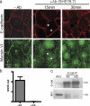
E-cadherin adhesion is necessary to recruit myosin VI to cell–cell contacts. (a) 48-h-old MCF7 cell monolayers were incubated in medium alone (−Ab) or in the presence of the E-cadherin function-blocking mAb SHE78-7 for 15–30 min. E-cadherin was detected using a mouse mAb against the cytoplasmic domain, whereas myosin VI was detected using myosin VI pAb. SHE78-7 abolished myosin VI staining at cadherin contacts within 15 min, before the integrity of the contacts was overtly disrupted (arrowheads). (b) Ratiometric analysis of myosin VI (MVI)/E-cadherin fluorescence intensity at cell–cell contacts in control cultures (−Ab) and cultures treated with SHE78-7 for 15 min (+Ab). Data are means ± SEM (error bars; n = 30–40 and are representative of four independent experiments). (c) E-cadherin immune complexes isolated from control monolayers (−Ab) or monolayers treated with SHE78-7 for 30 min (+Ab) were probed for myosin VI and E-cadherin.
Figure 3.
Figure 3.
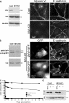
Myosin VI is necessary for E-cadherin adhesion and cohesive cell–cell contacts. MCF7 cells were transiently transfected with siRNA directed against human myosin VI (MVI-KD) or with control duplexes bearing a single mismatched base pair (Cont). (a) siRNA depletes myosin VI. Western analysis of myosin VI expression in cells 6 d after transfection. Loading controls were β-tubulin (Tub) and GAPDH. Indirect immunofluorescence microscopy showed that myosin VI was largely undetectable at E-cadherin contacts. Compared with control cells, E-cadherin staining in myosin VI KD cells was punctate and discontinuous. (b) Exogenous myosin VI rescues cell contact integrity in myosin VI KD cells. Myosin VI expression was reconstituted by the transient expression of porcine myosin VI tagged with EGFP (pMVI-GFP). Western blotting for myosin VI revealed both the transgene and endogenous myosin VI. Endogenous human myosin VI expression was reduced, but that of the transgene was not affected by the myosin VI siRNA. Linear, continuous E-cadherin staining was seen at contacts between cells expressing pMVI-GFP (arrowhead) compared with untransfected controls or cells expressing the myosin VI tail domain alone (pMVI–tail-GFP; arrowheads). (c) Depletion of myosin VI reduces E-cadherin–based adhesion. Adhesion of control MCF7 cells or myosin VI KD cells to hE/Fc-coated substrata was measured using laminar flow assays and was expressed as the percentage of cells that remained adherent to hE/Fc at various detachment flow rates. E-cadherin–deficient CHO cells were used as negative controls. Data are means ± SEM (error bars; n = 3) and are representative of four independent experiments. (d) Myosin VI KD does not affect the total or surface expression levels of E-cadherin. Surface expression of E-cadherin was measured using surface trypsin protection assays in control cells or in cells depleted of myosin VI. Cells were lysed before (WCL) or after trypsinization in the presence (+) or absence (−) of extracellular Ca2+. Total levels of E-cadherin were unaffected by myosin VI KD, and surface E-cadherin remained sensitive to trypsinization in the absence of Ca2+ (−) in both the control and myosin VI–depleted cells. β-Tubulin was the loading control.
Figure 4.
Figure 4.
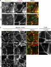
Myosin VI affects the perijunctional actin cytoskeleton. (a) Reorganization of the perijunctional actin cytoskeleton coincides with the accumulation of myosin VI. MCF7 cells cultured for 24 or 48 h were labeled for F-actin (phalloidin) and myosin VI. Whereas 24-h-old cultures showed loose perijunctional phalloidin staining, F-actin accumulated intensely at the cell–cell contacts in 48-h-old cultures. (b) Myosin VI depletion disrupts organization of the perijunctional actin cytoskeleton in 24- and 48-h-old cultures. Myosin VI KD cells showed loose perijunctional phalloidin staining compared with the dense peripheral staining seen in control (Cont) cells. E-cadherin was used to identify the cell–cell contacts between myosin VI KD cells. The dense perijunctional F-actin staining was restored in cells transiently expressing porcine myosin VI (KD + pMVI-GFP; asterisks).
Figure 5.
Figure 5.
Myosin VI supports the integrity of apical junctional complexes. Myosin VI KD and control MCF7 monolayers were immunolabeled for ZO-1, desmoplakin (DP), or vinculin to identify tight junctions, desmosomes, and the zonula adherens, respectively, and were imaged by spinning disc confocal microscopy. Vinculin was nearly totally lost from the apical cell–cell contacts in myosin VI KD cells, whereas vinculin staining in basal focal adhesions persisted.
Figure 6.
Figure 6.
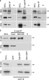
Myosin VI is necessary for E-cadherin and vinculin to interact. (a) Myosin VI, E-cadherin, and vinculin are found in a complex. Protein complexes were immunoprecipitated from 48-h-old MCF7 monolayers using either a myosin VI pAb (MVI-IP), E-cadherin pAb (E-cad-IP), or vinculin mAb (Vinc-IP). Western blots of immunoprecipitates and whole cell lysates (WCL) were probed for E-cadherin, myosin VI, and vinculin. Negative controls for immunoprecipitations were naive rabbit antisera for E-cadherin and myosin VI and anti-HA monoclonal antibody for vinculin (φ). (b) E-cadherin is unable to immunoprecipitate vinculin in the absence of myosin VI. Western blots of E-cadherin immunoprecipitates (E-cad IP) and whole cell lysates (WCL) from myosin VI KD or control cells were probed for E-cadherin or vinculin. Negative controls for immunoprecipitations (φ) were naive rabbit antisera. (c) The myosin VI tail coimmunoprecipitates E-cadherin and vinculin. Full-length p–myosin VI–GFP, p–myosin VI–tail-GFP, and GFP alone were transiently expressed in MCF7 cells. Western blots of anti-GFP immune complexes were probed for E-cadherin and vinculin. Blotting for GFP (on separate gels because of the disparate molecular weights) confirmed that the transgenes expressed polypeptides of predicted molecular weight and were immunoprecipitated to a similar level (not depicted).
Figure 7.
Figure 7.
Vinculin depletion disrupts the integrity of E-cadherin cell–cell contacts. MCF7 cells were transiently transfected with siRNA duplexes against human vinculin (Vinc-KD) or a control duplex with a 4-bp substitution (Cont). (a) Immunoblotting for vinculin and tubulin confirmed the efficient depletion of vinculin. (b) Immunofluorescence staining revealed that E-cadherin cell–cell contacts were discontinuous and serrated in vinculin KD cells compared with controls. (c) Vinculin is not necessary for myosin VI to coimmunoprecipitate E-cadherin. Myosin VI was immunoprecipitated from vinculin-depleted (Vinc-KD) or control cultures and immune complexes probed for both myosin VI and E-cadherin. Negative controls for immunoprecipitations were naive rabbit antisera (φ).
Figure 8.
Figure 8.
Membrane-targeted vinculin fragments rescue cell–cell integrity in myosin VI–depleted cells. (a) Schematic of constructs: full-length αE-catenin bearing a C-terminal T7 tag (α-catenin) and α-catenin/vinculin chimeric proteins in which the N-terminal 1–325 amino acid domain of α-catenin was fused to the N-terminal 1–823 head domain of vinculin (α-cat/vinHead) or the C-terminal 822–1,067 tail domain of vinculin (α-cat/vinTail). (b) Expression of vinculin constructs but not α-catenin rescues the integrity of E-cadherin cell–cell contacts in myosin VI–depleted cells. Myosin VI KD cells were transiently transfected with α-catenin–vinculin constructs or full-length α-catenin and were fixed and stained for E-cadherin. Transfected cells were identified by the T7 tag; nuclear staining in the T7-stained cells is background, as it was detected in nontransfected controls (not depicted). Linear E-cadherin contacts were frequently restored in cells expressing either α-catenin/vinculin construct (arrowheads). (c) Quantitation revealed that α-cat/vinTail did not rescue as frequently as αE/vinHead, whereas α-catenin was unable to rescue E-cadherin contact integrity to any comparable extent. Data are means ± SEM (error bars; n = 100 and are representative of five independent experiments).
Figure 9.
Figure 9.
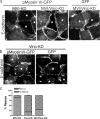
Vinculin is necessary for myosin VI to regulate cadherin contacts. (a) Myosin VI reconstitution requires vinculin to restore the integrity of E-cadherin contacts. Porcine myosin VI (p–myosin VI–GFP) or GFP alone (GFP) were transiently expressed in MCF7 cells transfected with siRNA for myosin VI alone (MVI-KD) or with siRNA for both myosin VI and vinculin (MVI/Vinc-KD). Cells were stained for E-cadherin and the GFP tag; cells expressing p–myosin VI–GFP or GFP are denoted by asterisks. P–myosin VI–GFP failed to restore linear cadherin contacts in myosin VI/vinculin double KD cells. (b) Overexpression of myosin VI does not rescue linear cadherin contacts in vinculin- depleted cells. P–myosin VI–GFP or GFP alone were transiently expressed in cells transfected with siRNA for vinculin. Samples were stained for E-cadherin, and cells expressing the transgene (asterisks) were identified by GFP. (c) Rescue of linear contacts by myosin VI. The ability of p–myosin VI–GFP to rescue linear cadherin contacts was assessed in cells depleted of myosin VI alone (MVI-KD), vinculin alone (Vinc-KD), and depleted of both myosin VI and vinculin (MVI/Vinc-KD). Contacts were scored based on whether they were linear (rescue) or discontinuous (no rescue); data are means ± SEM (error bars; n = 100 and are representative of five independent experiments).

Similar articles

Cited by

References

    1. Adams, C.L., Y.-T. Chen, S.J. Smith, and W.J. Nelson. 1998. Mechanisms of epithelial cell-cell adhesion and cell compaction revealed by high-resolution tracking of E-cadherin-green fluorescent protein. J. Cell Biol. 142:1105–1119. - PMC - PubMed
    1. Altman, D., H.L. Sweeney, and J.A. Spudich. 2004. The mechanism of myosin VI translocation and its load-induced anchoring. Cell. 116:737–749. - PubMed
    1. Aschenbrenner, L., T. Lee, and T. Hasson. 2003. Myo6 facilitates the translocation of endocytic vesicles from cell peripheries. Mol. Biol. Cell. 14:2728–2743. - PMC - PubMed
    1. Au, J.S., C. Puri, G. Ihrke, J. Kendrick-Jones, and F. Buss. 2007. Myosin VI is required for sorting of AP-1B-dependent cargo to the basolateral domain in polarized MDCK cells. J. Cell Biol. 177:103–114. - PMC - PubMed
    1. Bryant, D.M., and J.L. Stow. 2004. The ins and outs of E-cadherin trafficking. Trends Cell Biol. 14:427–434. - PubMed

Publication types

MeSH terms