. 2007 Aug 7;104(32):13010-5.
doi: 10.1073/pnas.0700970104. Epub 2007 Jul 30.

Assays of vacuole fusion resolve the stages of docking, lipid mixing, and content mixing

Affiliations

Assays of vacuole fusion resolve the stages of docking, lipid mixing, and content mixing

Youngsoo Jun et al. Proc Natl Acad Sci U S A. .

Abstract

Membrane fusion entails organelle docking and subsequent mixing of membrane bilayers and luminal compartments. We now present an in vitro assay of fusion, using yeast vacuoles bearing domains of either Fos or Jun fused to complementary halves of beta-lactamase. Upon fusion, these proteins associate to yield beta-lactamase activity. This assay complements the standard fusion assay (activation of pro-Pho8p in protease-deficient vacuoles by proteases from pho8Delta vacuoles). Both the beta-lactamase and pro-Pho8p activation assays of fusion show the same long kinetic delay between SNARE pairing and luminal compartment mixing. Lipid-mixing occurs rapidly after SNARE pairing but well before aqueous compartment mixing. These results support a model in which SNARE pairing leads to rapid hemifusion, followed by slow further lipid rearrangement and aqueous compartment mixing.


Conflict of interest statement

The authors declare no conflict of interest.

Figures
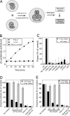
Fig. 1.
Fig. 1.
A vacuole fusion assay. (A) An in vitro vacuole fusion assay based on complementary reconstitution of β-lactamase. (B) Fusion-dependent β-lactamase activity formation over the 90-min time course. BJ3505-Fos-ω vacuoles, BJ3505-α-Jun vacuoles, or both were incubated at 27°C in fusion reactions. A portion of reactions was transferred to ice at indicated times and assayed for β-lactamase activity. One unit (U) of the β-lactamase activity is defined as 1 nmol of hydrolyzed nitrocefin per min from 12 μg of vacuole protein. (C) Comparison between the β-lactamase (black bars) and ALP (gray bars) fusion assays. Fusion reactions (see Materials and Methods) were incubated for 90 min on ice or at 27°C with indicated inhibitors. One reaction lacked ATP. Values were normalized to the reactions without inhibitors (3.78 ± 0.65 units for the ALP assay; 1.76 ± 0.45 units for the β-lactamase assay). Data represent mean ± SEM (n = 3). Inhibitors concentrations: affinity-purified anti-Sec17p (153 nM), affinity-purified anti-Sec18p (142 nM), anti-Vam3p IgG (444 nM), affinity-purified anti-Vps33p (155 nM), MED (10 μM), and U73122 (60 μM). (D and E) The β-lactamase fusion assay is less sensitive to microcystin-LR (MCLR) and GTPγS than the ALP assay. The β-lactamase (gray bars) and the ALP (black bars) fusion assays were performed in the presence of increasing concentrations of GTPγS (D) or MCLR (E) and antibodies to Sec17p where indicated. Values were normalized to the reactions without inhibitors (4.6 ± 0.38 units for the ALP assay; 1.74 ± 0.27 units for the β-lactamase assay). Data represent mean ± SEM (n = 3).
Fig. 2.
Fig. 2.
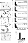
GTPγS allows fusion but prevents pro-ALP activation. Fusion reactions were performed under standard conditions but with 10 μg of vacuoles. After 90 min, aliquots were stained with 3.3 μM MDY-64 for fluorescent microscopy (A–F) or assayed for ALP activity (G). Images were acquired and analyzed by using ImageJ 1.36b and JMP 5.0.1a (SAS Institute). Vacuole diameters were measured from micrographs taken from random fields, and the surface areas of individual vacuoles were plotted as distribution histograms (F). A representative field for each condition is shown (Scale bars, 20 μm).
Fig. 3.
Fig. 3.
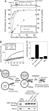
The R18-dequenching lipid mixing assay. (A) Vacuoles were labeled with R18, mixed with unlabeled vacuoles in lipid mixing assay reactions containing anti-Sec17p (154 nM), anti-Sec18p (142 nM), anti-Ypt7p (167 nM), Gdi1p (1.2 μM)/Gyp1–46p (5 μM), anti-Vam3p (444 nM), anti-Nyv1p (261 nM), MED (10 μM), 3sQ-SNARE (2 μM), recombinant Sec18p (63 nM), rVam7p (40 nM), or buffer only (No addition), and lipid mixing was monitored at 27°C. One reaction did not receive unlabeled acceptor vacuoles (open circles). (B) After 90 min, ALP activity was measured. (C and D) Vacuoles isolated from BJ3505-Fos-ω were labeled with R18, reisolated, and added to lipid mixing assay reactions containing unlabeled DKY6281 vacuoles (C) or unlabeled vacuoles from BJ3505-α-Jun (D). Lipid mixing (squares) was monitored by R18 dequenching under low Mg2+ condition at 27°C for 90 min with the indicated concentrations of GTPγS. After 90 min, the reactions were assayed for ALP activity (C; circles) or β-lactamase activity (D; circles). Values obtained without inhibitors [2.31 ± 0.46 units for the ALP assay coupled with the R18 assay (ΔF90min /FTX100 = 0.102 ± 0.047); 1.53 ± 0.07 units for the β-lactamase assay coupled with the R18 assay (ΔF90min /FTX100 = 0.201 ± 0.023)] were used as the 100% reference. Data represent mean ± SEM (n = 3).
Fig. 4.
Fig. 4.
Kinetics of lipid and content mixing. (A) Kinetic separation of SNARE complex assembly and lipid mixing from content mixing. ALP fusion reactions (1 μg of R18-labeled BJ3505 vacuoles and 5 μg of unlabeled DKY6281 vacuoles) contained affinity-purified anti-Sec17p (154 nM). Recombinant Vam7p (1 μM) was added after 20 min. A portion of each reaction was transferred to a microplate fluorometer and monitored for R18 dequenching at 27°C for 64 min. The remainder received anti-Vam3p IgG or were placed on ice at indicated times (−-0.5, 0.5, 1, 2, 4, 8, 16, 32, 48, or 64 min). ALP activity was measured after 64 min. Fusion values were normalized to reactions that were placed on ice at 64 min (3.48 ± 0.3). The relative fluorescence change ΔF64min/FTX100 (see Materials and Methods) at 64 min (0.124 ± 0.002) was the 100% reference for lipid mixing. Data represent mean ± SEM (n = 3). (B–D) MED allows trans-SNARE complex assembly but blocks lipid mixing and content mixing. Vacuoles isolated from the BJ3505-CBP-Vam3p nyv1Δ yeast strain were labeled with 150 μM R18, reisolated, and added to lipid mixing assay reactions containing unlabeled vacuoles isolated from BJ3505-CBP-Vam3p nyv1Δ and DKY6281 yeast strains. The lipid mixing assay reactions (480 μl) contain 10.7 μg of R18-labeled vacuoles and 85.3 μg of unlabeled vacuoles. Lipid mixing was monitored at 27°C without inhibitors, with Gdi1p (1.2 μM)/Gyp1–46p (5 μM) or with MED (10 μM) (B). After 60 min, a portion of reactions (30 μl) was withdrawn to assay ALP activity (C). Data represent mean ± SEM (n = 3). The remaining reactions (450 μl) were subjected to assay of trans-SNARE complexes (see Materials and Methods). The transassociation of CBP-Vam3p from the BJ3505-CBP-Vam3p nyv1Δ vacuoles with Nyv1p from the DKY6281 vacuoles is shown (D).

Similar articles

Cited by

References

    1. Cheever ML, Sato TK, de Beer T, Kutateladze TG, Emr SD, Overduin M. Nat Cell Biol. 2001;3:613–618. - PubMed
    1. Stroupe C, Collins KM, Fratti RA, Wickner W. EMBO J. 2006;25:1579–1589. - PMC - PubMed
    1. Kemble GW, Danieli T, White JM. Cell. 1994;76:383–391. - PubMed
    1. Melikyan GB, White JM, Cohen FS. J Cell Biol. 1995;131:679–691. - PMC - PubMed
    1. Chernomordik LV, Frolov VA, Leikina E, Bronk P, Zimmerberg J. J Cell Biol. 1998;140:1369–1382. - PMC - PubMed

Publication types

MeSH terms