. 2007 Jul 31;104(31):12603-9.
doi: 10.1073/pnas.0610755104. Epub 2007 Jul 30.

Single-molecule fluorescence spectroscopy in (bio)catalysis

Affiliations

Single-molecule fluorescence spectroscopy in (bio)catalysis

Maarten B J Roeffaers et al. Proc Natl Acad Sci U S A. .

Abstract

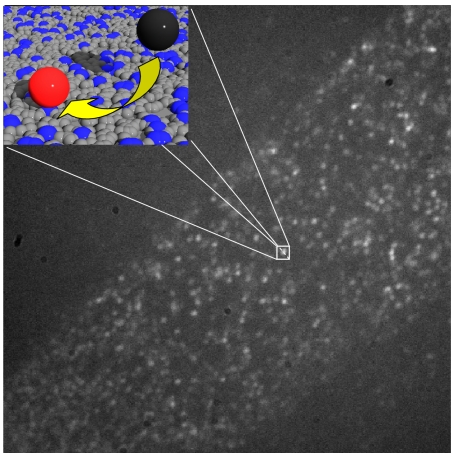
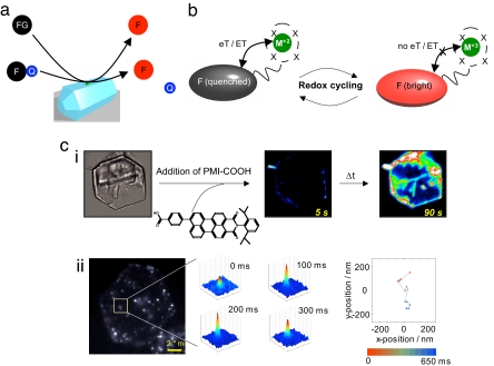
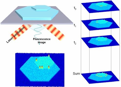
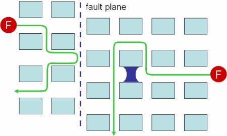
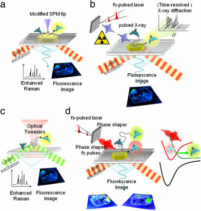
The ever-improving time and space resolution and molecular detection sensitivity of fluorescence microscopy offer unique opportunities to deepen our insights into the function of chemical and biological catalysts. Because single-molecule microscopy allows for counting the turnover events one by one, one can map the distribution of the catalytic activities of different sites in solid heterogeneous catalysts, or one can study time-dependent activity fluctuations of individual sites in enzymes or chemical catalysts. By experimentally monitoring individuals rather than populations, the origin of complex behavior, e.g., in kinetics or in deactivation processes, can be successfully elucidated. Recent progress of temporal and spatial resolution in single-molecule fluorescence microscopy is discussed in light of its impact on catalytic assays. Key concepts are illustrated regarding the use of fluorescent reporters in catalytic reactions. Future challenges comprising the integration of other techniques, such as diffraction, scanning probe, or vibrational methods in single-molecule fluorescence spectroscopy are suggested.


Conflict of interest statement

The authors declare no conflict of interest.

Figures

Fig. 1.
Fig. 1.
Stochastic nature of turnover events. This wide-field image was recorded during hydrolysis of fluorogenic fluorescein esters on a monolayer containing aminopropyl groups diluted by propyl groups. The amino group density is much higher than the spatial resolution of the fluorescence microscope. However, because each group is only sporadically active, the individual turnover events can be observed as isolated bright spots. (Inset) Density of aminopropyl groups (blue) versus propyl groups (gray) and the conversion of a nonfluorescent substrate (black) into a fluorescent product (red).
Fig. 2.
Fig. 2.
Disorder in catalysts. (a) Interconverting enzyme conformations with different activities/selectivities. (b) Some of the Os(VIII) species that can osmylate olefins during asymmetric dihydroxylation. (c) Within a population of crystals, one can distinguish different crystal habitus (1 versus 2), different degrees of intergrowth (3), and on the crystal planes of individual crystals, different sites (4).
Fig. 3.
Fig. 3.
Fluorescence-based visualization of catalytic sites and events. (a) A fluorescent product (F) is formed by transformation of a fluorogenic reactant (FG) or by cleavage of a covalently bound quencher (Q). (b) Cycling of a metal catalyst (M) between two redox states causes quenching and dequenching of a fluorescent reporter (F). (c) Mapping of basic sites on a layered double hydroxide crystal with acid probes: (i) Imaging of time-dependent sorption of a perylene monoimide carboxylic acid. Even at the ensemble level differences between crystal faces can be distinguished (ii). At the single-molecule level, the probe hops between individual basic sites.
Fig. 4.
Fig. 4.
High-resolution reconstruction of the active-site distribution in a catalyst. The individual frames show stochastic turnover events; superimposition of consecutively recorded frames yields the high-resolution image as in photoactivation high-resolution light microscopy (45).
Fig. 5.
Fig. 5.
Tracking of single fluorescent molecules (F) diffusing in porous materials allows finding of local obstructions or fault planes.
Fig. 6.
Fig. 6.
Combination of techniques that can be envisioned for in situ characterization of catalytic systems. (a) SMFS and tip-enhanced Raman spectroscopy. Catalytic conversion can be followed by fluorescence while chemical characterization of the active site can be obtained simultaneously. (b) SMFS and (time-resolved) x-ray experiments. This combination allows for simultaneous mapping of catalytic activity and crystallographic data. (c) Optical trapping for immobilizing small catalyst particles in solution. This approach minimizes diffusion resistances. (d) SMFS combined with phase-shaped femtosecond pulses can control the outcome of the catalytic reaction.

Similar articles

Cited by

References

    1. de Levie R. J Chem Educ. 2000;77:771–774.
    1. Xie XS, Lu HP. J Biol Chem. 1999;274:15967–15970. - PubMed
    1. Engelkamp H, Hatzakis NS, Hofkens J, De Schryver FC, Nolte RJM, Rowan AE. Chem Commun. 2006;9:935–940. - PubMed
    1. Smiley RD, Hammes GG. Chem Rev. 2006;106:3080–3094. - PubMed
    1. Craig DB, Arriaga EA, Wong JCY, Lu H, Dovichi NJ. J Am Chem Soc. 1996;118:5245–5253.

Publication types

LinkOut - more resources