. 2008 Feb;19(2):861-8.
doi: 10.1007/s10856-007-0166-6. Epub 2007 Jul 31.

The behavior of osteoblast-like cells on various substrates with functional blocking of integrin-beta1 and integrin-beta3

Affiliations

The behavior of osteoblast-like cells on various substrates with functional blocking of integrin-beta1 and integrin-beta3

M C Siebers et al. J Mater Sci Mater Med. 2008 Feb.

Abstract

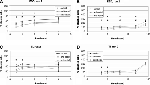
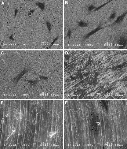
This study was designed to examine the influence of integrin subunit-beta1 and subunit-beta3 on the behavior of primary osteoblast-like cells, cultured on calcium phosphate (CaP)-coated and non coated titanium (Ti). Osteoblast-like cells were incubated with specific monoclonal antibodies against integrin-beta1 and integrin-beta3 to block the integrin function. Subsequently, cells were seeded on Ti discs, either non coated or provided with a 2 microm carbonated hydroxyapatite coating using Electrostatic Spray Deposition. Results showed that on CaP coatings, cellular attachment was decreased after a pre-treatment with either anti-integrin-beta1 or anti-integrin-beta3 antibodies. On Ti, cell adhesion was only slightly affected after a pre-treatment with anti-integrin-beta3 antibodies. Scanning electron microscopy showed that on both types of substrate, cellular morphology was not changed after a pre-treatment with either antibody. With quantitative PCR, it was shown for both substrates that mRNA expression of integrin-beta1 was increased after a pre-treatment with either anti-integrin-beta1 or anti-integrin-beta3 antibodies. Furthermore, after a pre-treatment with either antibody, mRNA expression of integrin-beta3 and ALP was decreased, on both types of substrate. In conclusion, osteoblast-like cells have the ability to compensate to great extent for the blocking strategy as applied here. Still, integrin-beta1 and beta3 seem to play different roles in attachment, proliferation, and differentiation of osteoblast-like cells, and responses on CaP-coated substrates differ to non coated Ti. Furthermore, the influence on ALP expression suggests involvement of both integrin subunits in signal transduction for cellular differentiation.


Figures

Fig. 1
Fig. 1
Cellular attachment in the first run. (A) and (B) CaP-coated substrates derived with Electrostatic Spray Deposition (ESD); (C) and (D) non-coated Ti. * indicates a significant difference between the control group and the experimental group with the pre-treatment with anti-integrin-β1 antibodies. indicates a significant difference between the control group and the experimental group that was pre-treated with anti-integrin-β3 antibodies
Fig. 2
Fig. 2
Cellular attachment in the second run. (A) and (B) CaP-coated substrates derived with Electrostatic Spray Deposition (ESD); (C) and (D) non-coated Ti. * indicates a significant difference between the control group and the experimental group with the pre-treatment with anti-integrin-β1 antibodies. # indicates a significant difference between the control group and the experimental group that was pre-treated with anti-integrin-β3 antibodies
Fig. 3
Fig. 3
Scanning electron micrographs of osteoblast-like cells on CaP-coated (A, B, and C) substrates, and non-coated (D, E, and F) Ti substrates after 1 day of culture. There were no morphological differences between the control group (A and D), or the experimental groups that were either pre-treated with anti-integrin-β1 antibodies (B and E), or anti-integrin-β3 antibodies (C and F)

References

    1. {'text': '', 'ref_index': 1, 'ids': [{'type': 'DOI', 'value': '10.1034/j.1600-0501.1998.090407.x', 'is_inner': False, 'url': 'https://doi.org/10.1034/j.1600-0501.1998.090407.x'}, {'type': 'PubMed', 'value': '9760901', 'is_inner': True, 'url': 'https://pubmed.ncbi.nlm.nih.gov/9760901/'}]}
    2. S. VERCAIGNE, J. G. C. WOLKE, I. NAERT and J. A. JANSEN, Clin. Oral Impl. Res.9 (1998) 261 - PubMed
    1. {'text': '', 'ref_index': 1, 'ids': [{'type': 'DOI', 'value': '10.1034/j.1600-0501.2000.011004314.x', 'is_inner': False, 'url': 'https://doi.org/10.1034/j.1600-0501.2000.011004314.x'}, {'type': 'PubMed', 'value': '11168224', 'is_inner': True, 'url': 'https://pubmed.ncbi.nlm.nih.gov/11168224/'}]}
    2. S. VERCAIGNE, J. G. C. WOLKE, I. NAERT and J. A. JANSEN, Clin. Oral Impl. Res.11 (2000) 314 - PubMed
    1. {'text': '', 'ref_index': 1, 'ids': [{'type': 'DOI', 'value': '10.1006/cbir.1996.0110', 'is_inner': False, 'url': 'https://doi.org/10.1006/cbir.1996.0110'}, {'type': 'PubMed', 'value': '9046103', 'is_inner': True, 'url': 'https://pubmed.ncbi.nlm.nih.gov/9046103/'}]}
    2. M. CASTOLDI, M. PISTONE, C. CARUSO, A. PUDDU, C. FILANTI, D. PICCINI, C .TACCHETTI and P. MANDUCA, Cell Biol. Int.21 (1997) 7 - PubMed
    1. {'text': '', 'ref_index': 1, 'ids': [{'type': 'DOI', 'value': '10.1016/S0142-9612(99)00242-2', 'is_inner': False, 'url': 'https://doi.org/10.1016/s0142-9612(99)00242-2'}, {'type': 'PubMed', 'value': '10711964', 'is_inner': True, 'url': 'https://pubmed.ncbi.nlm.nih.gov/10711964/'}]}
    2. K. ANSELME, Biomaterials21 (2000) 667 - PubMed
    1. {'text': '', 'ref_index': 1, 'ids': [{'type': 'DOI', 'value': '10.1016/S8756-3282(99)00007-1', 'is_inner': False, 'url': 'https://doi.org/10.1016/s8756-3282(99)00007-1'}, {'type': 'PubMed', 'value': '10221544', 'is_inner': True, 'url': 'https://pubmed.ncbi.nlm.nih.gov/10221544/'}]}
    2. G. B. SCHNEIDER, S. W. WHITSON and L. F. COOPER, Bone24 (1999) 321 - PubMed

Publication types

LinkOut - more resources