. 2007 Oct 15;407(2):313-20.
doi: 10.1042/BJ20070794.

Timed interactions between viral and cellular replication factors during the initiation of SV40 in vitro DNA replication

Affiliations

Timed interactions between viral and cellular replication factors during the initiation of SV40 in vitro DNA replication

Poonam Taneja et al. Biochem J. .

Abstract

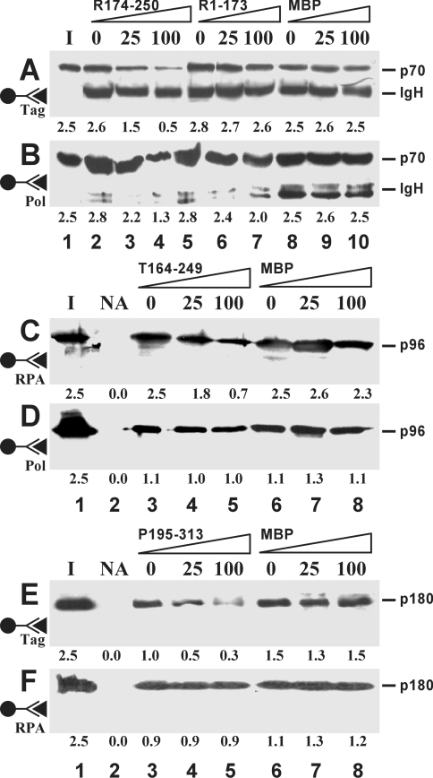
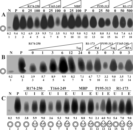
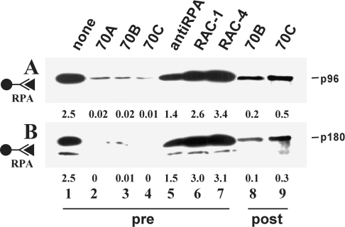
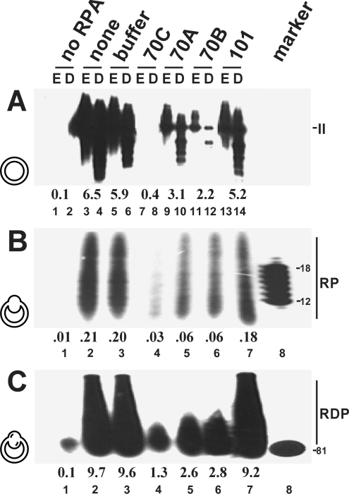
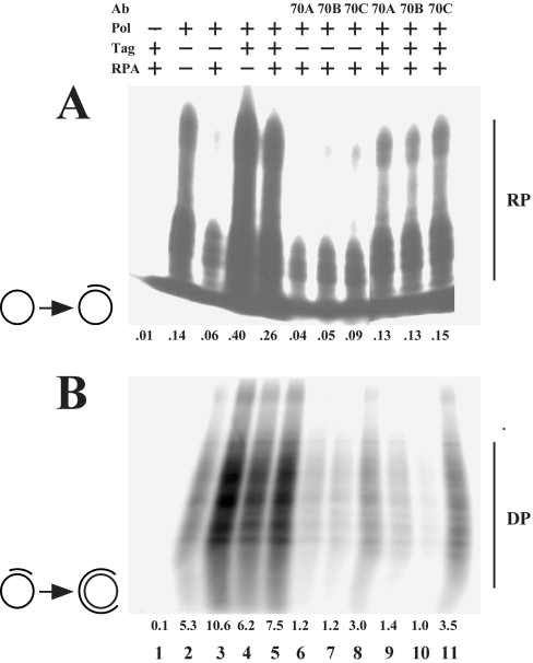
The initiation of SV40 (simian virus 40) DNA replication requires the co-operative interactions between the viral Tag (large T-antigen), RPA (replication protein A) and Pol (DNA polymerase alpha-primase) on the template DNA. Binding interfaces mapped on these enzymes and expressed as peptides competed with the mutual interactions of the native proteins. Prevention of the genuine interactions was accomplished only prior to the primer synthesis step and blocked the assembly of a productive initiation complex. Once the complex was engaged in the synthesis of an RNA primer and its extension, the interfering effects of the peptides ceased, suggesting a stable association of the replication factors during the initiation phase. Specific antibodies were still able to disrupt preformed interactions and inhibited primer synthesis and extension activities, underlining the crucial role of specific protein-protein contacts during the entire initiation process.


Figures

Figure 1
Figure 1. Inhibition of protein–protein interactions by peptides
Pairwise co-precipitations were performed between Tag and RPA (A, C), Pol and RPA (B, F), and Pol and Tag (D, E), each at 25 pmol. The immobilized bait proteins are indicated by the antibody symbol on the left. Co-precipitated target proteins Tag (p96), Pol (p180 subunit) and RPA (p70 subunit) were revealed by Western blotting using the ECL® detection system. Peptides were present at a 25- and 100-fold molar excess over the target protein or were omitted (0). The numbers below each lane represent the average amount (in pmol) of co-precipitated protein as determined by densitometric scanning from five independent experiments. The position of the heavy chain of immunoglobulin (IgH) is indicated when it was detected due to cross-reactivity of the secondary antibodies. Lane I, one-tenth of the input material of soluble target proteins; lane NA, no antibody added.
Figure 2
Figure 2. Influence of peptides on the monopolymerase system
(A) Titration of peptides. Increasing amounts of indicated peptides displayed as fold molar excess over 6.25 pmol of Tag were titrated into the reaction at the onset of monopolymerase reactions. (B) Back-titration of replication factors. Replication factors were added back at the indicated amounts at the onset of the reactions. Molar excess of peptides over Tag was 100-fold (for R1-173, 500-fold). (C) Kinetics of inhibition. Indicated peptides were added to the reaction mixture at a 100-fold (500-fold for R1-173) molar excess over Tag before unwinding (U), primer synthesis (I) and primer elongation (E). The different template configurations present at the time at which the peptides were administered were under-wound (U), primed (I) and primer-extended (E) plasmid DNAs, which are symbolized for each reaction. Lanes N and P represent negative and positive controls in the absence and presence of Tag. The numbers above lane numbers denote the amount of nucleotides incorporated (in pmol).
Figure 3
Figure 3. Influence of p70-specific monoclonal antibodies on protein–protein interactions
H6-RPA was linked to beads via anti-His antibodies. The resin was incubated with the indicated antibodies before (pre) or after (post) addition of the other replication factors. Anti-RPA refers to a polyclonal serum. Co-precipitated proteins were detected by Western blotting using the ECL® detection system. The positions of Tag (p96) and the p180 subunit of Pol are indicated. Lane 1 contained one-tenth of the input material of the soluble target protein. Numbers below each lane indicate the amounts of co-precipitated proteins (in pmol) as determined by densitometry.
Figure 4
Figure 4. Influence of p70-specific antibodies on replication-related activities of RPA
Antibodies were tested in SV40 DNA replication (A), primer synthesis (B) and primer extension (C) assays. In (A), each DNA was restricted with EcoRI alone (E, odd-numbered lanes) or double- restricted by EcoRI–DpnI (D, even-numbered lanes). The numbers below the lanes indicate the amounts (in pmol) of incorporated dNMPs (A, C) or rNMPs (B). Lane 1 contained no protein. The positions of the products of each reaction, linear double-stranded DNA (form II), RNA primers (RP) and RNA–DNA primers (RDP), are indicated. Markers contained oligo(dT)8-12 or an 81-base-pair fragment. Antibodies were present at the onset of the reaction (A) or before the unwinding (B) or primer synthesis (C) steps. The template configurations present at the time at which antibodies were administered were super-coiled, under-wound and primed plasmid DNAs and are represented by symbols for each reaction.
Figure 5
Figure 5. Influence of antibodies on primer synthesis and extension using natural ssDNA templates
Primer synthesis (A) and primer extension (B) reactions were carried out using an un-primed or primed single-stranded circular DNA respectively as the templates, which are indicated by symbols for each reaction. The presence of any of the replication proteins Tag, Pol and RPA are indicated by ‘+’, their absence by ‘–’. Antibodies were added as specified (lanes 6–11). The numbers below each lane indicate the incorporated amounts (in pmol) of rNMPs (A) and dNMPs (B). RNA (RP) and DNA (DP) products are indicated.

Similar articles

Cited by

References

    1. Kelly T. J. SV40 DNA replication. J. Biol. Chem. 1988;263:17889–17892. - PubMed
    1. Stenlund A. Initiation of DNA replication: lessons from viral initiator proteins. Nat. Rev. Mol. Cell Biol. 2003;4:777–785. - PubMed
    1. Simmons D. T. SV40 large T antigen functions in DNA replication and transformation. Adv. Virus Res. 2000;55:75–134. - PubMed
    1. Waga S., Bauer G., Stillman B. Reconstitution of complete SV40 DNA replication with purified replication factors. J. Biol. Chem. 1994;269:10923–10934. - PubMed
    1. Stauffer M. E., Chazin W. J. Structural mechanisms of DNA replication, repair, and recombination. J. Biol. Chem. 2004;279:30915–30918. - PubMed

Publication types

MeSH terms

LinkOut - more resources