. 2007 Sep;100(3):483-96.
doi: 10.1093/aob/mcm141. Epub 2007 Jul 30.

Formation of specialized propagules resistant to desiccation and cryopreservation in the threatened moss Ditrichum plumbicola (Ditrichales, Bryopsida)

Affiliations

Formation of specialized propagules resistant to desiccation and cryopreservation in the threatened moss Ditrichum plumbicola (Ditrichales, Bryopsida)

J K Rowntree et al. Ann Bot. 2007 Sep.

Abstract

Background and aims: Successful cryopreservation of bryophytes is linked to intrinsic desiccation tolerance and survival can be enhanced by pre-treatment with abscisic acid (ABA) and sucrose. The pioneer moss Ditrichum plumbicola is naturally subjected to desiccation in the field but showed unexpectedly low survival of cryopreservation, as well as a poor response to pre-treatment. The effects of the cryopreservation protocol on protonemata of D. plumbicola were investigated in order to explore possible relationships between the production in vitro of cryopreservation-tolerant asexual propagules and the reproductive biology of D. plumbicola in nature.

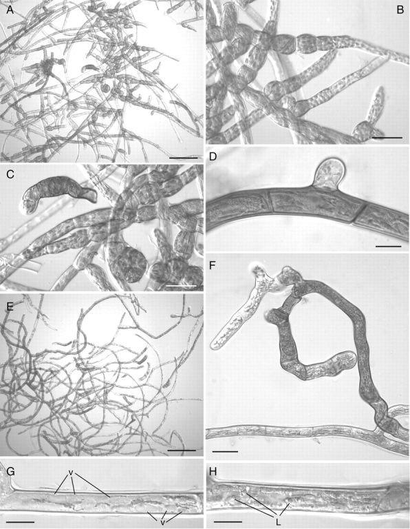
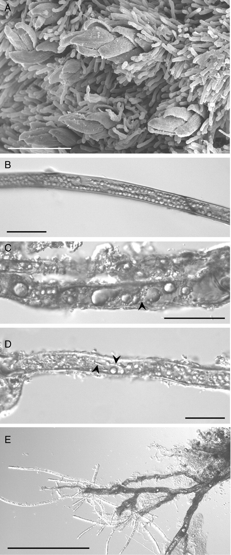
Methods: Protonemata were prepared for cryopreservation using a four-step protocol involving encapsulation in sodium alginate, pre-treatment for 2 weeks with ABA and sucrose, desiccation for 6 h and rapid freezing in liquid nitrogen. After each stage, protonemata were prepared for light and electron microscopy and growth on standard medium was monitored. Further samples were prepared for light and electron microscopy at intervals over a 24-h period following removal from liquid nitrogen and re-hydration.

Key results: Pre-treatment with ABA and sucrose caused dramatic changes to the protonemata. Growth was arrested and propagules induced with pronounced morphological and cytological changes. Most cells died, but those that survived were characterized by thick, deeply pigmented walls, numerous small vacuoles and lipid droplets in their cytoplasm. Desiccation and cryopreservation elicited no dramatic cytological changes. Cells returned to their pre-dehydration and cryopreservation state within 2 h of re-hydration and/or removal from liquid nitrogen. Regeneration was normal once the ABA/sucrose stimulus was removed.

Conclusions: The ABA/sucrose pre-treatment induced the formation of highly desiccation- and cryopreservation-tolerant propagules from the protonemata of D. plumbicola. This parallels behaviour in the wild, where highly desiccation-tolerant rhizoids function as perennating organs allowing the moss to endure extreme environmental conditions. An involvement of endogenous ABA in the desiccation tolerance of D. plumbicola is suggested.


Figures

F<sc>ig</sc>. 1.
Fig. 1.
Growth data (mean diameter in mm) at week 5 for control (1), encapsulation (2), ABA/sucrose (3), desiccation (4) and cryopreservation (5) treatments of Ditrichum plumbicola. Error bars = s.e., n = 50. Encapsulation treatment shows significantly reduced growth compared with the control (P < 0·001). ABA/sucrose, desiccation and cryopreservation treatments show significantly reduced growth compared with both the control and encapsulation treatments (P < 0·001), but not each other.
F<sc>ig</sc>. 2.
Fig. 2.
Ditrichum plumbicola protonema after 5 weeks of treatment: (A) control, (B) encapsulation, (C) ABA/sucrose, (D) desiccation and (E) cryopreservation treatments.
F<sc>ig</sc>. 3.
Fig. 3.
Light micrographs of cultured protonemata of Ditrichum plumbicola – controls and encapsulated material. (A–D) Controls: (A) typical chloronema cell with ovoid peripheral chloroplasts; (B) typical caulonema cell with elongated, longitudinally aligned plastids; (C) typical rhizoid with thick brown-pigmented walls, minute elongated plastids and spindle-shaped nucleus (n); (D) dedifferentiation of aerial chloronemal cells into thin-walled brood cells after 2–3 weeks in culture. (E–H) Encapsulated material: (E, F) typical chloronema (E) and caulonema (F) cells indistinguishable from the controls; (G) thin-walled brood cells on chloronema filaments after 2–3 weeks in culture; (H) 1-μm section of chloronema cell – note the large central vacuole and the peripherally located plastids (p) and nucleus. Scale bars: D, E, G = 50 µm; A–C, F = 20 µm; H = 10 µm.
F<sc>ig</sc>. 4.
Fig. 4.
Light micrographs of sucrose/ABA-treated protonemata: (A) the majority of the cells are dead; those that survive are characterized by heavily pigmented walls; (B) detail of dead cells showing complete disruption of cell contents; (C) heavily pigmented filament with dense cytoplasm and with non-pigmented apical cell (arrowed); (D) detail of non-pigmented apical cell – the arrow indicates incomplete cross wall; (E) lipid-laden chloronemal cells with non-pigmented thick walls; (F–H) encapsulated, sucrose/ABA-treated cells after 2 weeks from transfer to standard growth medium. The protonemal system remains virtually unchanged except for increased brood cell development (F) and detachment of the same (arrowed) from the parent filament (H). Scale bars: A, F = 200 µm; B, C = 100 µm; E, H = 50 µm; D, G = 20 µm.
F<sc>ig</sc>. 5.
Fig. 5.
Transmission electron micrographs of sucrose/ABA-treated protonemal cells before and after dehydration. (A–E) Fully hydrated cells: (A) cell with starch-filled plastid and with thick, multi-layered cell wall – note the numerous projections of the cell wall innermost layer; (B) detail of cell wall projection consisting of loosely fibrillar material; (C) incomplete cross wall (arrowed) in apical cell – note the thin, homogeneous cell wall (cw); (D) cell packed with small vacuoles – also note the centrally located round nucleus (n) and the starch-filled plastids; (E) cytoplasm containing mitochondria (m) with dense matrix and saccate cristae and vacuoles (v) with electron-opaque content. (F–H) Dehydrated cells: (F) ovoid plastids with small starch grains (arrowed) and with an intact, undulating thylakoid system; (G) nucleus (n) with conspicuous blocks of condensed chromatin surrounded by small vacuoles; (H) heterogeneous cell wall (cw) with irregular internal projections. Scale bars: D = 5 µm; A–C, E–G = 2 µm; H = 1 µm.
F<sc>ig</sc>. 6.
Fig. 6.
Mean diameter (mm) of Ditrichum plumbicola protonema grown for 5 weeks on control (open circles, continuous line), 5 % sucrose (open triangles, continuous line), 10 µM ABA (closed circles, dashed line) and 5 % sucrose/10 µM ABA (closed triangle, dashed line) media. Error bars are s.e., n = 59. Significance values from two-way ANOVA at week 5 are shown.
F<sc>ig</sc>. 7.
Fig. 7.
Light micrographs of ABA- and sucrose-treated protonemal cells. (A–D) Treatment for 2 weeks with 10 µM ABA: (A) most filaments are thick-walled and deeply pigmented; (B) dedifferentiation of chloronema filaments into spherical brood cells; (C) caulonema filament with thick pigmented walls – note the short cells with dense cytoplasm and the very short side branch. (E–H) Treatment for 2 weeks with 5 % sucrose: (E) general aspect showing thick-walled pigmented filaments as in (A) but no brood cells; (F) detail of thick-walled pigmented filament – note the thin-walled non-pigmented apical cells in the side branch; (G, H) caulonema cells with numerous small vacuoles (v) (G) and abundant lipid droplets (L) (H) in their cytoplasm. Scale bars: A, E = 200 µm; B, C, F = 50 µm; D, G, H = 20 µm.
F<sc>ig</sc>. 8.
Fig. 8.
Light micrographs of frozen and thawed protonemal cells. (A) Frozen cells mounted in immersion oil, the cells are flattened and the discoidal plastids are distributed throughout the cell lumen. (B–E) Frozen cells 1 h after thawing: (B, C) elongate plastids (p) in cytoplasm packed with lipid droplets (L) and small vacuoles – note the ovoid nucleus (n) with prominent nucleolus and no evidence of condensed chromatin in (C); (D) apical cells with round nuclei and less dense cytoplasm; (E) apical cell with incomplete cross wall (arrowed). (F) Cells kept in the original alginate strip 2 weeks after thawing – the cytology remains unchanged; (G, H) Production of normal filaments 2 d (G) and 4 d (H) after removing the thawed protonema from the alginate strips. Scale bars: G, H = 200 µm; A–F = 20 µm.
F<sc>ig</sc>. 9.
Fig. 9.
Transmission electron micrographs of frozen and thawed protonemal cells. (A–E) Frozen cells: (A) elongated plastids and nucleus with condensed chromatin – note the numerous lipid droplets and the small vacuoles in the cell lumen; (B) plastids with intact thylakoids and small starch grains; (C) rounded mitochondria with thin, parallel-sided cristae; (D) ovoid plastids with starch grains in an apical cell. (E) Short segments of ER (arrowed) remaining in the lumen of an apical cell (cw, cell wall). (F–H) Frozen cell 1–4 h after thawing: (F) the cytoplasm is packed with small vacuoles and numerous lipid droplets; (G) profiles of tubular ER (arrowed) are scattered in the cytoplasm amongst the mitochondria and the plastids, which now contain numerous starch grains, especially those of the apical cells (H). cw, Cell wall. Scale bars: A, F = 5 µm; B, D, E, G, H = 2 µm; C = 1 µm.
F<sc>ig</sc>. 10.
Fig. 10.
Ditrichum plumbicola from the wild: (A) scanning electron micrograph of mature colony showing D. plumbicola gametophores overgrown by the perennial protonemata of Pogonatum aloides; (B–D) light micrographs of herbarium (B) and freshly collected (C and D) material showing thick-walled rhizoids packed with lipid droplets of various sizes and only occasional starch grains (C) – in (D) note the minute plastids (arrowed) aligned along cytoplasmic strands like those seen in culture (see Fig. 4C); (E) normal protonemal system regenerating from > 1-year-old herbarium material. Scale bars: E = 100 µm; B–D = 50 µm; A = 20 µm.

References

    1. Alpert P. Constraints of tolerance: why are desiccation-tolerant organisms so small or rare? Journal of Experimental Biology. 2006;209:1575–1584. - PubMed
    1. Arts T. Rhizoidal tubers and protonemal gemmae in European Ditrichum species. Journal of Bryology. 1994;18:43–61.
    1. Beckett RP. ABA-induced tolerance to ion leakage during rehydration following desiccation in the moss Atrichum androgynum. Plant Growth Regulation. 2001;35:131–135.
    1. Beckett RP, Csintalan Z, Tuba Z. ABA treatment increases both the desiccation tolerance of photosynthesis, and nonphotochemical quenching in the moss Atrichum undulatum. Plant Ecology. 2000;151:65–71.
    1. Benson EE. Cryopreservation. In: Benson EE, editor. Plant conservation biotechnology. London: Taylor & Francis; 1999. pp. 83–95.

Publication types