. 2007 Aug 7;104(32):13022-7.
doi: 10.1073/pnas.0704059104. Epub 2007 Jul 31.

Intrinsic disorder in the C-terminal domain of the Shaker voltage-activated K+ channel modulates its interaction with scaffold proteins

Affiliations

Intrinsic disorder in the C-terminal domain of the Shaker voltage-activated K+ channel modulates its interaction with scaffold proteins

Elhanan Magidovich et al. Proc Natl Acad Sci U S A. .

Abstract

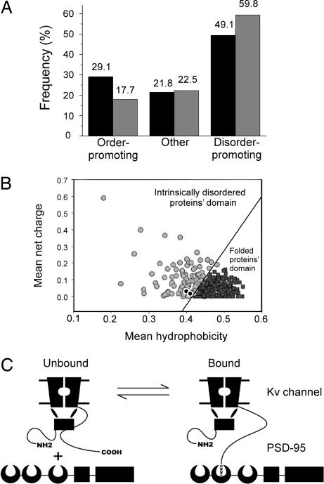
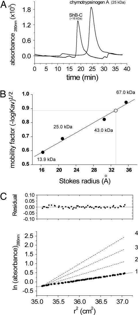
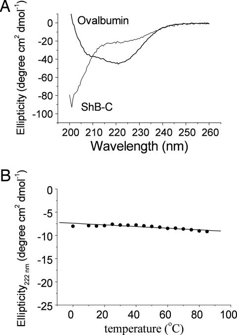
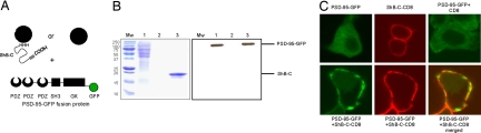
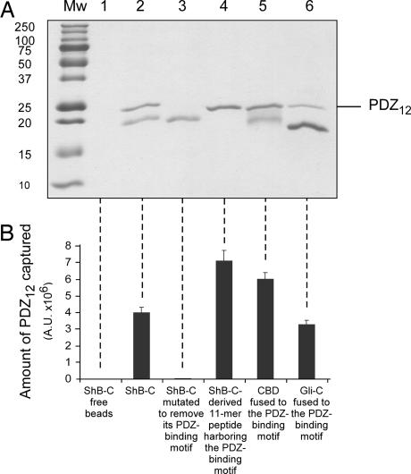
The interaction of membrane-embedded voltage-activated potassium channels (Kv) with intracellular scaffold proteins, such as the postsynaptic density 95 (PSD-95) protein, is mediated by the channel C-terminal segment. This interaction underlies Kv channel clustering at unique membrane sites and is important for the proper assembly and functioning of the synapse. In the current study, we address the molecular mechanism underlying Kv/PSD-95 interaction. We provide experimental evidence, based on hydrodynamic and spectroscopic analyses, indicating that the isolated C-terminal segment of the archetypical Shaker Kv channel (ShB-C) is a random coil, suggesting that ShB-C belongs to the recently defined class of intrinsically disordered proteins. We show that isolated ShB-C is still able to bind its scaffold protein partner and support protein clustering in vivo, indicating that unfoldedness is compatible with ShB-C activity. Pulldown experiments involving C-terminal chains differing in flexibility or length further demonstrate that intrinsic disorder in the C-terminal segment of the Shaker channel modulates its interaction with the PSD-95 protein. Our results thus suggest that the C-terminal domain of the Shaker Kv channel behaves as an entropic chain and support a "fishing rod" molecular mechanism for Kv channel binding to scaffold proteins. The importance of intrinsically disordered protein segments to the complex processes of synapse assembly, maintenance, and function is discussed.


Conflict of interest statement

The authors declare no conflict of interest.

Figures

Fig. 1.
Fig. 1.
The C-terminal tail domain of the Shaker Kv channel is predicted to be intrinsically disordered. (A) Comparison of the relative order- and disorder-promoting amino acid content across the entire Drosophila proteome (black bars) and in ShB-C (gray bars). (B) The mean net charge and mean hydrophobicity values of the Shaker and Kv 1.2 channels (black circles) lie within the intrinsically disordered protein domain of Uversky's phase-space diagram. The solid line is an empirical linear regression that defines the boundary between folded (black squares) and intrinsically disordered (gray circles) proteins (adapted with permission from ref. 11). (C) A fishing rod mechanism for Kv channel binding to scaffold proteins. A voltage-gated K+ channel interacts with the PSD-95 scaffold protein upon binding of the C-terminal PDZ-binding motif hook, tethered to an intrinsically disordered extended chain. The moon-shape, box, and rectangular shapes represent the PDZ, SH3, and guanylate kinase domains of the PSD-95 protein, respectively.
Fig. 2.
Fig. 2.
Hydrodynamic analyses reveal an ShB-C structure with low compactness. (A) Size-exclusion chromatography elution profiles of ShB-C and chymotrypsinogen A, monitored at 280 nm. (B) Mobility [(−logKav)1/2; see Materials and Methods] vs. Stokes radius plot, used for Stokes radius determination of ShB-C, based on analytical size-exclusion chromatography of standard monomeric molecular weight markers. The open circle corresponds to ShB-C. The black circles correspond to molecular weight markers, as indicated. (C) Equilibrium sedimentation of ShB-C at 5°C and 19,000 rpm. Representative data are plotted as ln (absorbance) against the square of the radius from the axis of rotation. The slope is proportional to the molecular mass (see SI Text). Dashed lines with increasing slopes indicate calculated values for monomeric ShB-C (1), dimeric (2), trimeric (3), and tetrameric (4) forms of ShB-C. The data are consistent with a monomeric model for ShB-C, as judged by the residuals plot.
Fig. 3.
Fig. 3.
CD spectroscopic analysis reveals an extended conformation for ShB-C. (A) Comparison of the far-UV CD spectra of ovalbumin and ShB-C (0.5 mg/ml), obtained at room temperature (25°C). The CD spectrum of ShB-C was essentially similar at 90°C (not shown). (B) Temperature dependence of the molar ellipticity of ShB-C (0.125 mg/ml), followed at 222 nm.
Fig. 4.
Fig. 4.
Isolated ShB-C binds its PSD-95 scaffold protein partner and supports protein clustering in vivo. (A) Schematic depiction of the experimental setup used for the batch pulldown assay. Ni2+-NTA bead-bound ShB-C protein containing the PDZ-binding motif at its C terminus (gray rectangular box) served as bait for the capture of the modular PSD-95 partner protein. (B) SDS/PAGE (Left) and Western blot (Right) analyses of eluted fractions of a pulldown experiment demonstrating the molecular interaction between ShB-C and PSD-95. Left lane corresponds to protein molecular weight markers. Lane 1 corresponds to a crude extract of Drosophila S2 Schneider cells transfected to express the PSD-95-GFP fusion protein. Lanes 2 and 3 correspond to the Drosophila crude extract incubated with ShB-C-free or -bound beads, respectively. Western blot analysis of the same gel shown (Right) was performed by using anti-GFP primary antibodies (see SI Text). (C) Confocal microscopic analysis of Drosophila S2 Schneider cells expressing PSD-95-GFP and/or ShB-C fused to the CD8 membrane-targeting sequence, either separately (Upper) or together (Lower).
Fig. 5.
Fig. 5.
The intrinsically disordered nature of ShB-C modulates its interaction with the PSD-95 scaffold protein. (A) SDS/PAGE analysis of eluted fractions of pulldown experiments using different ShB-C-derived chains as bait for the PDZ12 protein (see Materials and Methods). Lanes 1–5 correspond to pulldown experiments using (i) beads alone or beads containing the following chains as bait: (ii) ShB-C, (iii) the ShB-C protein mutated in its PDZ-binding motif, (iv) an ShB-C mutant lacking the entire C-terminal intrinsically disordered segment apart from the 11 last amino acids, (v) an ShB-C mutant protein in which the intrinsically disordered segment but not the PDZ-binding motif was replaced by the folded cellulose-binding domain, or (vi) an ShB-C mutant protein in which the complete intrinsically disordered segment but not the PDZ-binding motif was replaced by the intrinsically disordered C-terminal domain of gliotactin (Gli-C). (B) Densitometric analysis of the results presented in A. Each reported value represents an average of eight independent measurements. Differences in the amount of captured PDZ12 in each category, relative to ShB-C, were all found to be statistically significant, as judged by two-sided Student's t test. Because multiple comparisons of ShB-C to the other protein chains are involved, we followed Bonferroni's correction and used a more stringent criteria to reject the null hypothesis that the two compared groups are identical, based on a P value <1%.

References

    1. Sigworth FJ. Q Rev Biophys. 1994;27:1–40. - PubMed
    1. Yellen G. Q Rev Biophys. 1998;31:239–295. - PubMed
    1. Bezanilla F. Physiol Rev. 2000;80:555–592. - PubMed
    1. Hille B. Ion Channels of Excitable Membranes. Sunderland, MA: Sinauer; 2001.
    1. Tejedor FJ, Bokhari A, Rogero O, Gorczyca M, Zhang J, Kim E, Sheng M, Budnik V. J Neurosci. 1997;17:152–159. - PMC - PubMed

Publication types

MeSH terms

Substances

LinkOut - more resources