

Anatomy of the round window and hook region of the cochlea with implications for cochlear implantation and other endocochlear surgical procedures
- PMID: 17667773
- PMCID: PMC2556227
- DOI: 10.1097/mao.0b013e3180577949
Anatomy of the round window and hook region of the cochlea with implications for cochlear implantation and other endocochlear surgical procedures
Abstract
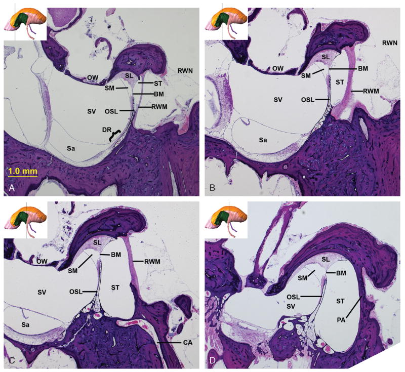
Hypothesis: The goal of this study was to create a three-dimensional model of the anatomy of the hook region to identify the optimal site for cochleostomy in cochlear implant surgery.
Background: The anatomy of the hook region is complex, and spatial relationships can be difficult to evaluate using two-dimensional histological slides or cadaveric temporal bones.
Methods: The right temporal bone of a 14-year-old adolescent boy was used to create a three-dimensional model. Sections containing the round window membrane (RWM) and surrounding cochlear structures were stained, digitized, and imported into a general purpose three-dimensional rendering and analysis software program (Amira, version 4.1). Three-dimensional models of the RWM, basilar membrane, osseous spiral lamina, spiral ligament, cochlear aqueduct, inferior cochlea vein, scala media, ductus reuniens, scala vestibuli, scala tympani, and surrounding bone were generated. The relationship between these structures and the RWM and adjacent otic capsule was evaluated. Histological sections from a different temporal bone were also analyzed. This temporal bone was sectioned in a plane perpendicular to the axis corresponding to the surgical view of the RWM, seen through the facial recess.
Results: The anteroinferior margin of the RWM or adjacent otic capsule was identified as the site for a cochleostomy that will avoid damage to critical cochlear structures and allow implantation directly into the scala tympani. The model can be downloaded from: https://research.meei.harvard.edu/otopathology/3dmodels.
Conclusion: This three-dimensional model has implications for surgical procedures to the inner ear that aim to minimize insertional trauma.
Figures


References
-
- Gantz BJ, Turner C. Combining acoustic and electrical speech processing: Iowa/Nucleus hybrid implant. Acta Otolaryngol. 2004;124:344–7. - PubMed
-
- Gantz BJ, Turner C, Gfeller KE, Lowder MW. Preservation of hearing in cochlear implant surgery: advantages of combined electrical and acoustical speech processing. Laryngoscope. 2005;115:796–802. - PubMed
-
- Gstoettner W, Kiefer J, Baumgartner WD, Pok S, Peters S, Adunka O. Hearing preservation in cochlear implantation for electric acoustic stimulation. Acta Otolaryngol. 2004;124:348–52. - PubMed
-
- James C, Albegger K, Battmer R, et al. Preservation of residual hearing with cochlear implantation: how and why. Acta Otolaryngol. 2005;125:481–91. - PubMed
-
- Eshraghi AA, Yang NW, Balkany TJ. Comparative study of cochlear damage with three perimodiolar electrode designs. Laryngoscope. 2003;113:415–9. - PubMed
Publication types
MeSH terms
Grants and funding
LinkOut - more resources
Full Text Sources
