. 2007 Dec;15(12):2132-9.
doi: 10.1038/sj.mt.6300275. Epub 2007 Jul 31.

Intranuclear trafficking of episomal DNA is transcription-dependent

Affiliations

Intranuclear trafficking of episomal DNA is transcription-dependent

Joshua Z Gasiorowski et al. Mol Ther. 2007 Dec.

Abstract

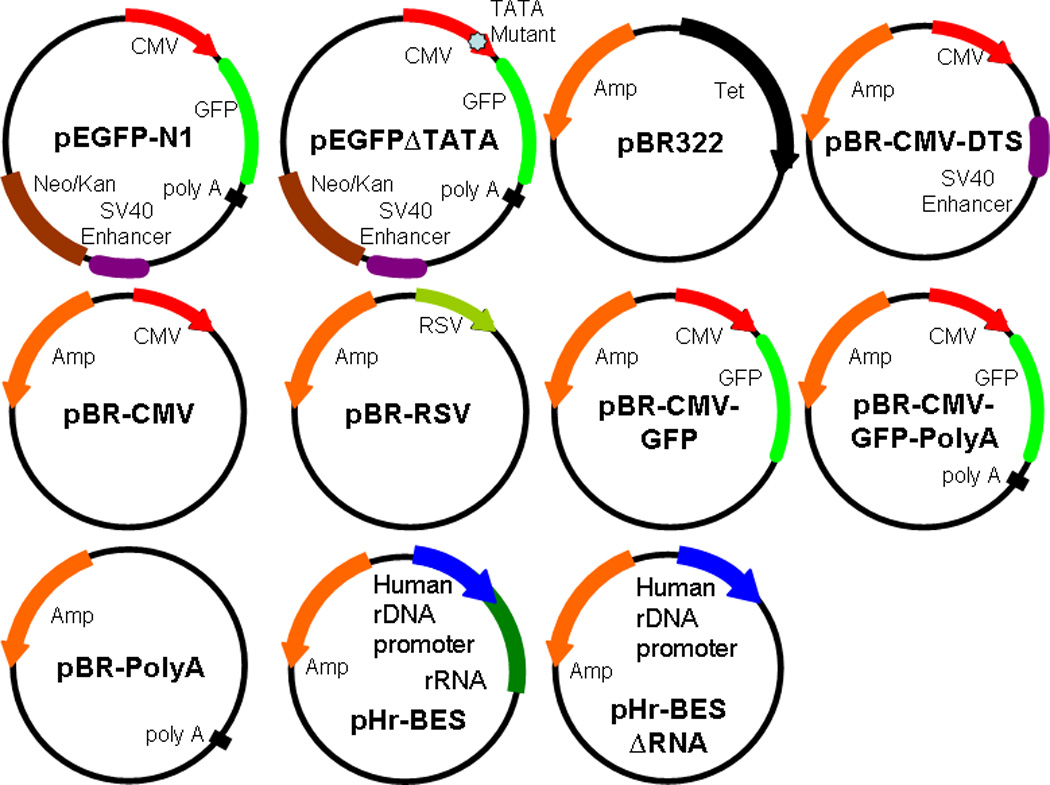
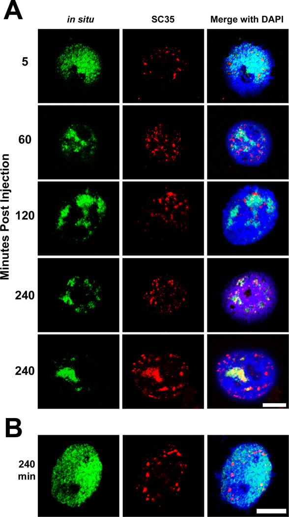
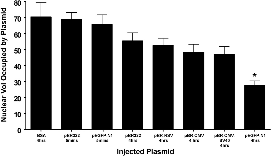
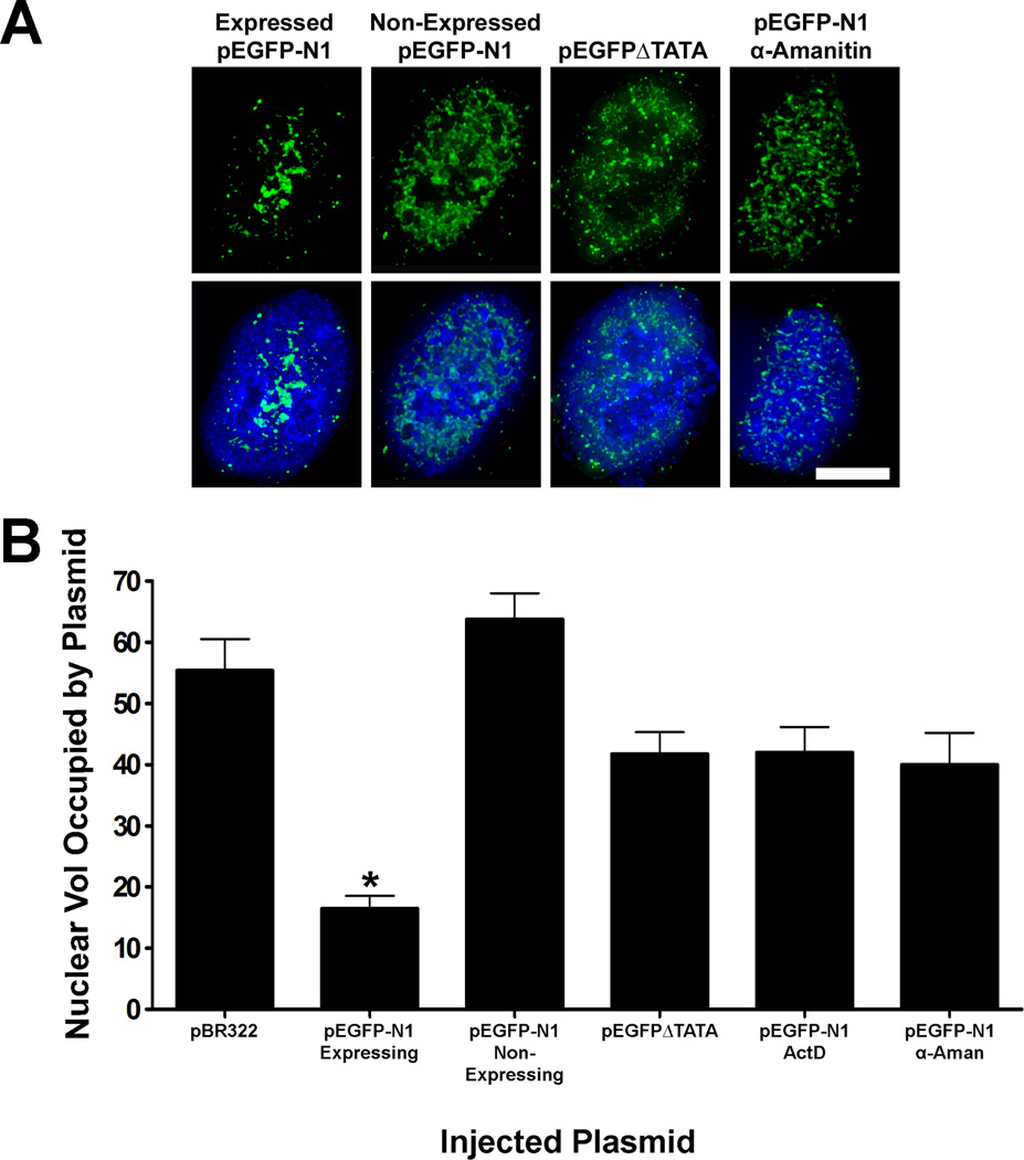
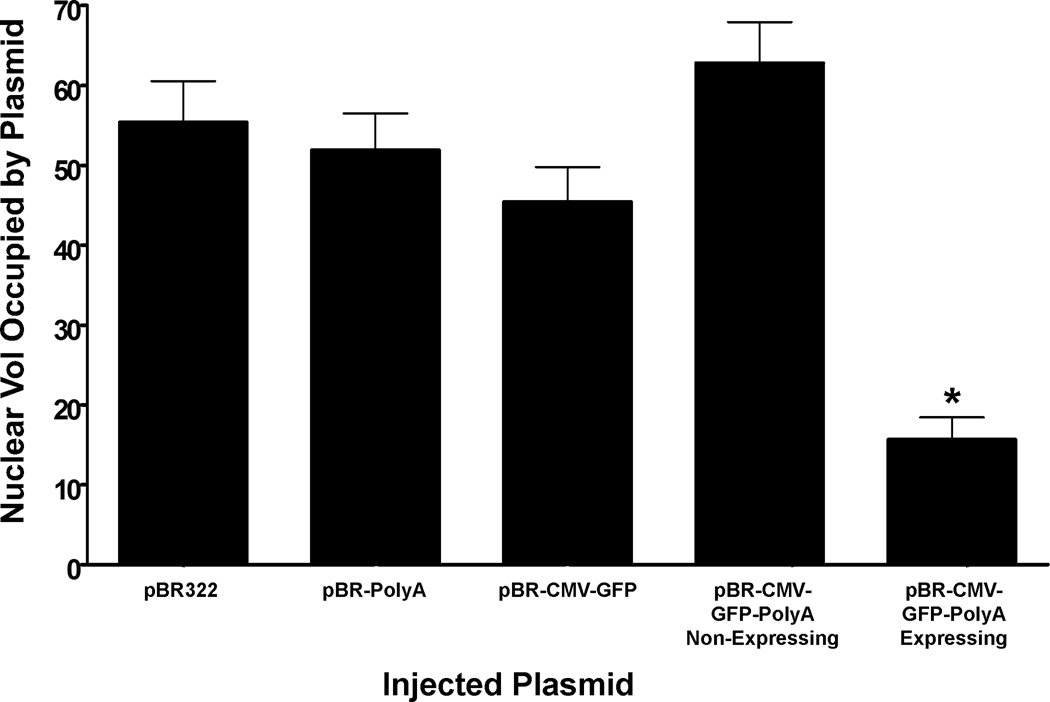
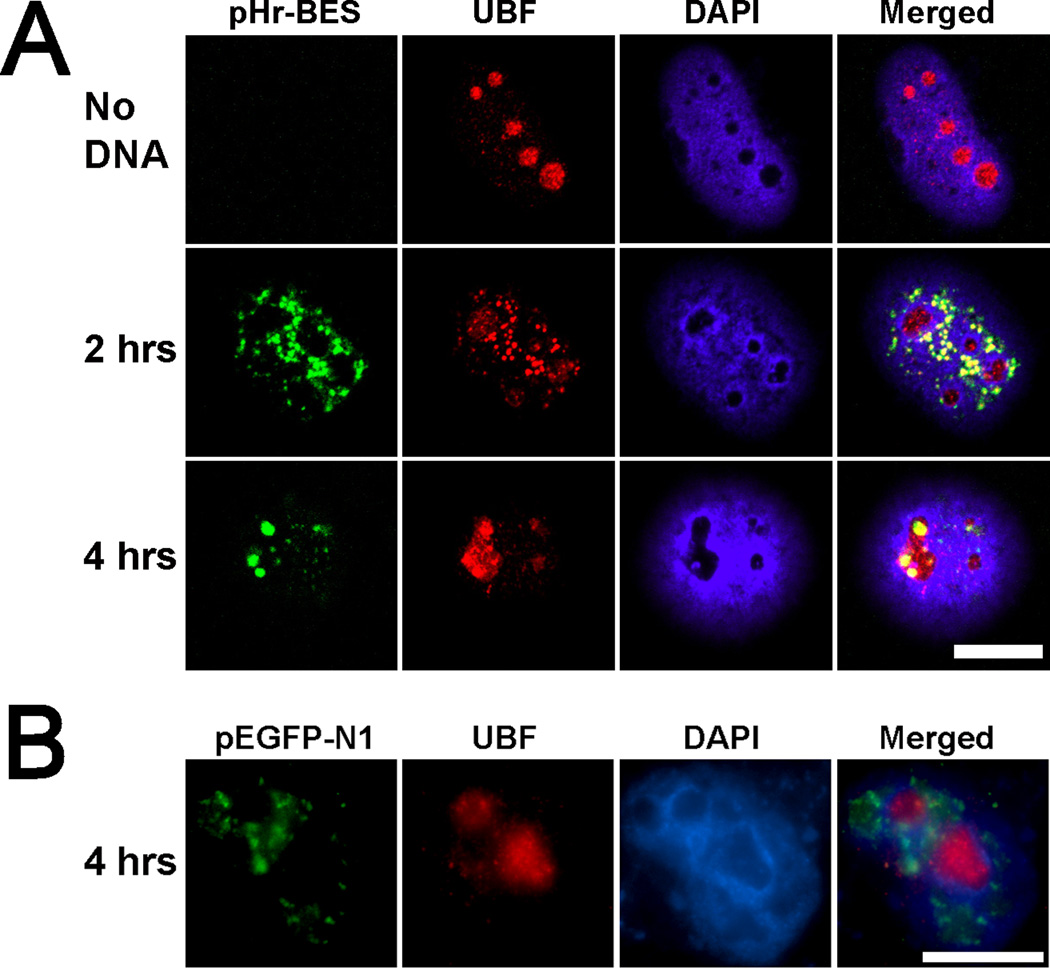
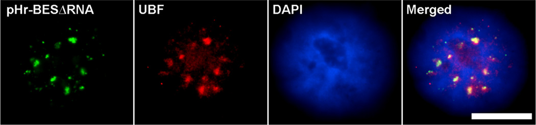
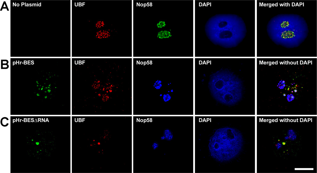
Our aim is to characterize the poorly understood mechanisms that influence episomal transgene expression within the nucleus. We found that plasmid DNA micro-injected directly into a nucleus moves into a speckled pattern and occupies less nuclear volume than bovine serum albumin (BSA) or other inert molecules after 4 hours. In addition, plasmids that contain eukaryotic regulatory sequences and actively transcribe transgenes condense into a few select areas of the nucleoplasm and occupy less nuclear volume than bacterial vectors. This suggests that episomal DNA moves in a sequence and transcription-dependent manner. We have also found that plasmids traffic to specific subnuclear domains depending on their sequence. Our experiments show that plasmids with polymerase II regulatory elements will target to nuclear spliceosome regions, while plasmids with the polymerase I promoter often traffic into nucleoli. Further elucidation of intranuclear plasmid trafficking behavior may lead to a better understanding of gene expression, and thereby help to improve basic laboratory techniques and clinical gene therapies.


Figures

Figure 1
Figure 1. Plasmid maps
All of the plasmids used have the pEGFP-N1 (Clontech) or pBR322 backbone. pEGFP∆TATA contains a mutant TATA box. All of the pBR plasmids have self-explained eukaryotic regulatory elements cloned into the pBR322 backbone. pHr-BES has a pBR322 backbone and contains the human ribosomal DNA promoter and encodes the first 700 base pairs of the 45S pre-ribosomal RNA. pHr-BES∆RNA is the same as pHr-BES but has the encoded pre-ribosomal RNA sequence deleted.
Figure 2
Figure 2. Time course of microinjected plasmids
(A) The pEGFP-N1 plasmid was microinjected directly into TC7 nuclei and detected by fluorescence in situ hybridization and confocal microscopy between 5 and 240 minutes after microinjection (green). Colocalization with the spliceosome factor, SC35, was also determined by immunofluorescence (red). Nuclear DNA was visualized with DAPI (blue). Two representative cells are shown for the 240 minute time point to illustrate co-localization with SC35 speckles. (B) Nuclei were microinjected with pBR322 and plasmids were detected four hours later via in situ hybridization as in A. After four hours, the pBR322 plasmid shows little, if any intranuclear plasmid movement. All panels are representative images of over 150 injected and imaged cells for each time point and plasmid. Bar = 10 µm.
Figure 3
Figure 3. Percentage of nuclear volume occupied by microinjected plasmids
Different plasmids were individually injected into TC7 nuclei and detected by in situ hybridization. Z-stacks of injected nuclei were deconvolved and the voxels of plasmid and DAPI signal were measured. The graph displays the averages of several microinjection experiments for each condition. BSA does not seem to traffic to any specific spot within the nucleus and becomes very diffuse. Compared to pEGFP-N1 5 minutes post-injection, pBR322 and pBR-CMV-DTS all occupy similar nuclear volumes. However, pEGFP-N1 plasmids 4 hours post-injection move into more condensed subnuclear regions and occupy significantly less nuclear volume than all other conditions (p<0.01). Each bar represents the average ± SEM of all in situ positive nuclei from several experiments, and 50–150 nuclei were injected for each condition per experiment.
Figure 4
Figure 4. Transcription capability influences intranuclear plasmid movement
(A) Extended focus deconvolved images show that cells expressing GFP have a nuclear pEGFP-N1 pattern that is condensed while cells that do not express GFP have plasmid patterns that are more spread out throughout the entire nucleus. (B) The average plasmid occupied nuclear volume from several experiments for each condition is represented in the graph. Cells that actively express GFP have episomal plasmids that occupy an average of 16% nuclear volume, and is significantly different (p<0.0001) from non-expressing pEGFP-N1, pEGFP∆TATA, or pEGFP-N1 nuclei treated with either actinomycin D or α-amanitin. Each bar represents the average ± SEM of all in situ positive nuclei from several experiments, and 50–150 nuclei were injected for each condition per experiment (p<0.0001).
Figure 5
Figure 5. The poly-adenylation signal is required for transcription mediated intranuclear plasmid movement
At 4 hours post-injection, cells that actively express GFP from pBR-CMV-GFP-PolyA have episomal plasmids that occupy an average of 15.6% nuclear volume, and are significantly different (p<0.0001) from pBR322, pBR-PolyA, pBR-CMV-GFP, or non-expressing pBR-CMV-GFP-PolyA. Each bar represents the average ± SEM of all in situ positive nuclei from several experiments, and 50–150 nuclei were injected for each condition per experiment (p<0.0001).
Figure 6
Figure 6. A plasmid with ribosomal DNA sequences traffics into nucleoli after 4 hours
(A) The ribosomal transcription factor, UBF (red), is normally found within the nucleoli of uninjected cells (No DNA). pHr-BES, containing the human pol I promoter and encoded rRNA, was injected into HeLa nuclei and observed 2 and 4 hours later. Two hours post-injection, the pHr-BES in situ signal (green) colocalizes with UBF in the DAPI-stained nucleoplasm (blue). However, 4 hours after injection, the plasmid has moved into nucleoli and still remains colocalized with UBF. (B) pEGFP-N1 is not found in nucleoli and does not colocalize with UBF 4 hours post injection. Bar = 10 µm.
Figure 7
Figure 7. Plasmid trafficking into nucleoli is dependent on rRNA transcription
When pHr-BES∆RNA, which does not encode rRNA sequences, is injected into HeLa cell nuclei, it is organized into speckles and colocalizes with transcription factor UBF after 4 hours, but it fails to traffic into nucleolar space. Bar = 10 µm
Figure 8
Figure 8. rRNA processing factors are only recruited to plasmids with transcribed rRNA sequences
(A) UBF and the rRNA processing factor Nop58 are found in the nucleoli of uninjected HeLa cells. (B) pHr-BES colocalizes with UBF and Nop58 in the nucleoplasm two hours post nuclear injection. C) pHr-BES∆RNA colocalizes with UBF, but Nop58 remains nucleolar and does not colocalize with the plasmid 2 hours post nuclear injection.

Similar articles

Cited by

References

    1. Verschure PJ. Chromosome organization and gene control: it is difficult to see the picture when you are inside the frame. J Cell Biochem. 2006;99:23–34. - PubMed
    1. Shav-Tal Y, Darzacq X, Singer RH. Gene expression within a dynamic nuclear landscape. Embo J. 2006;25:3469–3479. - PMC - PubMed
    1. Jackson DA, Juranek S, Lipps HJ. Designing nonviral vectors for efficient gene transfer and long-term gene expression. Mol Ther. 2006;14:613–626. - PubMed
    1. Jenke AC, Stehle IM, Herrmann F, Eisenberger T, Baiker A, Bode J, Fackelmayer FO, Lipps HJ. Nuclear scaffold/matrix attached region modules linked to a transcription unit are sufficient for replication and maintenance of a mammalian episome. Proc Natl Acad Sci U S A. 2004;101:11322–11327. - PMC - PubMed
    1. Jenke BH, Fetzer CP, Stehle IM, Jonsson F, Fackelmayer FO, Conradt H, Bode J, Lipps HJ. An episomally replicating vector binds to the nuclear matrix protein SAF-A in vivo. EMBO Rep. 2002;3:349–354. - PMC - PubMed

Publication types

LinkOut - more resources