. 2007 Oct;72(8):965-76.
doi: 10.1038/sj.ki.5002456. Epub 2007 Aug 1.

Advanced glycation endproducts induce podocyte apoptosis by activation of the FOXO4 transcription factor

Affiliations

Advanced glycation endproducts induce podocyte apoptosis by activation of the FOXO4 transcription factor

P Y Chuang et al. Kidney Int. 2007 Oct.

Abstract

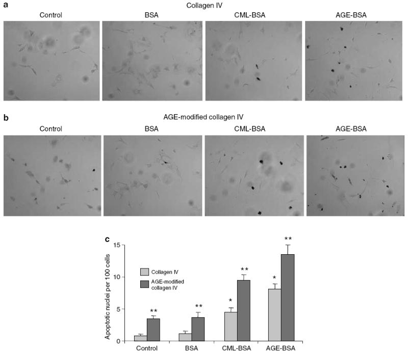
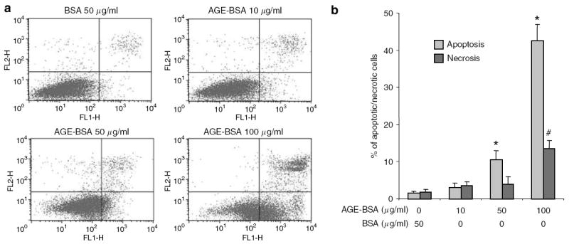
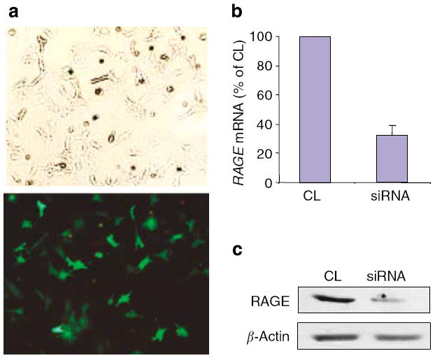
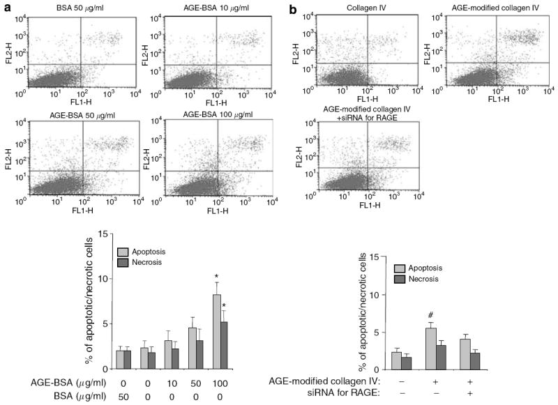
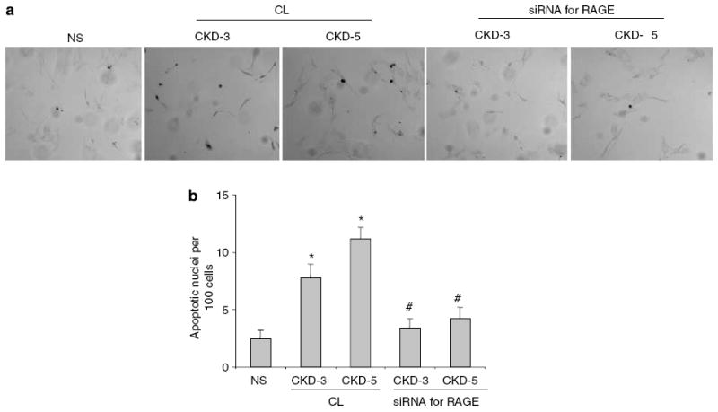
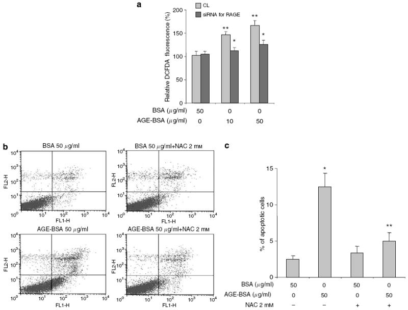
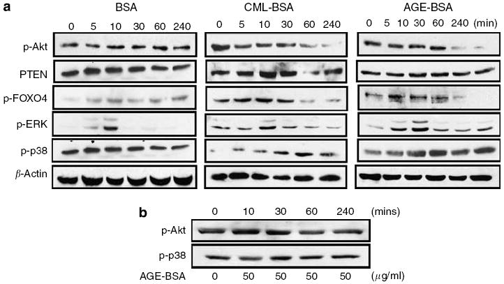
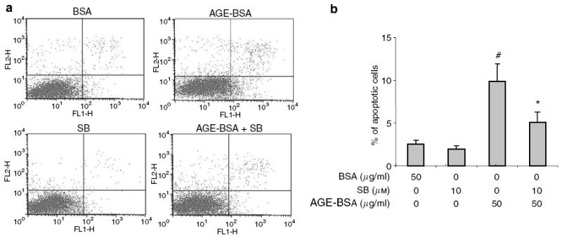
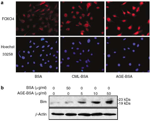
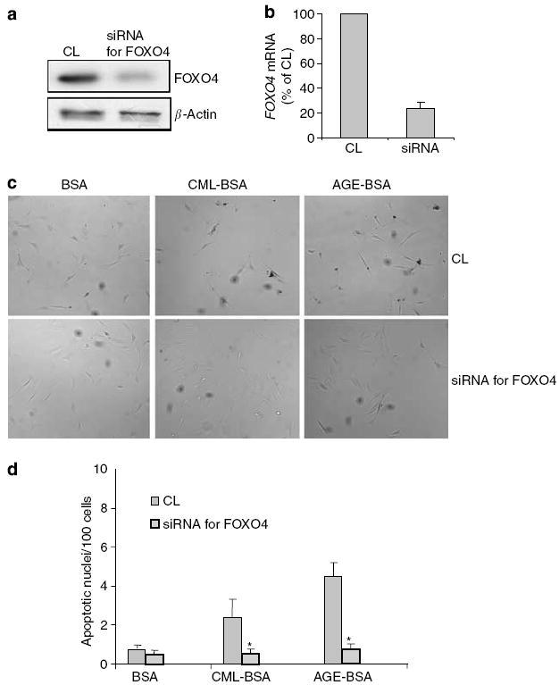
Advanced glycation endproducts (AGEs) and a receptor for AGEs (RAGE) have been linked in the pathogenesis of diabetic nephropathy. RAGE is usually localized to podocytes and is increased in diabetes. RAGE activation increases reactive oxygen species production, which mediates hyperglycemia-induced podocyte apoptosis in early diabetic nephropathy. Here, we examined the interaction of AGE and RAGE on podocyte apoptosis. When we exposed murine cultured podocytes to bovine serum albumin (BSA) that was modified by AGEs or to carboxymethyl-lysine BSA, more apoptosis was found when compared with unmodified BSA. Similarly, more podocytes underwent detachment and apoptosis when cultured on AGE-modified collagen IV than on native collagen IV. AGEs isolated from sera of patients with chronic kidney disease also caused apoptosis of podocytes. Apoptosis was diminished by small interference RNA (siRNA) for RAGE in podocytes exposed to AGE-BSA, but not to AGE-modified collagen IV. Both AGE- and carboxymethyl-lysine modified-BSA activated p38MAP kinase and inhibition of this kinase reduced the apoptotic effect of AGE-BSA. Exposure to AGE-BSA was associated with Akt dephosphorylation and FOXO4 transcriptional activation leading to an increase in the expression of an effector protein of apoptosis, Bim. siRNA for FOXO4 abolished AGE-BSA-induced apoptosis of podocytes. Our study suggests that an AGE-RAGE interaction contributes to podocyte apoptosis by activation of the FOXO4 transcription factor.


Figures

Figure 1
Figure 1. Podocyte apoptosis with soluble (CML-BSA and AGE-BSA) and matrix-bound AGE (AGE-modified collagen IV) exposures
Differentiated podocytes cultured on coverslips coated with either collagen IV or AGE-modified collagen IV were exposed to 50 μg/ml of BSA, AGE-BSA, or CML-BSA for overnight. Apoptosis of podocytes was determined by TUNEL staining. (a, b) Representative photographs of TUNEL staining are presented. (c) Bar graph representing the means±s.e.m. of the number of apoptotic nuclei per 100 cells in five fields at original magnification x 400. *P<0.01 as compared with BSA exposure on collagen-IV-coated coverslips (n =4). **P<0.01 as compared with corresponding collagen-IV-coated coverslips (n =4).
Figure 2
Figure 2. Podocytes undergo apoptosis after AGE-BSA exposure in a dose-dependent manner
Apoptosis and necrosis were quantified by FACS after Annexin V-FITC and PI labeling. (a) Representative FACS data for podocytes treated with BSA and AGE-BSA. The abscissa and ordinate represent the fluorescence intensity of Annexin V-FITC and PI, respectively. (b) Bar graph represents the mean percentages of apoptosis and necrosis±s.e.m. (n =3). *P<0.05 versus apoptosis of BSA 50 μg/ml. #P<0.05 versus necrosis of BSA 50 μg/ml.
Figure 3
Figure 3. Targeted gene knockdown of RAGE with siRNA
Transient transfection of podocytes with a siRNA for RAGE or the miRIDIAN microRNA Mimic negative control sequence (CL) was carried out using the Amaxa Nucleofection Device. Transfection efficiency was measured by the percentage of cells expressing GFP after transfection with pmaxGFP™. (a) Representative photographs of pmaxGFP™-transfected podocytes under light (top) and epifluorescence (bottom) microscopy. Effects of siRNA for RAGE and CL on RAGE mRNA level (b) and protein expression (c) were analyzed by real-time polymerase chain reaction and Western blotting, respectively. Representative data from three experiments are shown.
Figure 4
Figure 4. Knockdown of RAGE in podocytes mitigates the apoptotic effect of AGE-BSA, but not AGE-modified collagen IV
Apoptosis and necrosis were determined by FACS. Representative FACS data and bar graph summarizing the FACS results are shown. (a) Summary of FACS for apoptosis and necrosis of podocytes transfected with the siRNA for RAGE and treated with either BSA or AGE-BSA are shown. *P<0.05 versus BSA 50 μg/ml (n =3). (b) Apoptosis of podocytes transfected with the siRNA for RAGE and cultured on AGE-modified collagen IV. #P<0.05 versus the percentage of apoptosis in the other two groups (n =3).
Figure 5
Figure 5. AGEs isolated from sera of CKD patients induce podocyte apoptosis
The effect of AGEs isolated from sera of patients with stages 3 and 5 CKD-3 and CKD-5 on apoptosis was compared with those of normal subjects (NS). (a) Representative fields of TUNEL at original magnification x 400. (b) Podocytes were transfected with either a negative control oligonucleotide (CL) or the siRNA for RAGE. The mean percentages of apoptotic cells after exposure to AGEs from groups CKD-3 and CKD-5 are presented in the bar graph. *P<0.01 versus NS. (n =3) #P<0.01 versus the corresponding group treated with CL (n =3).
Figure 6
Figure 6. ROS production and apoptosis induced by AGE-BSA are diminished by a ROS scavenger, NAC
ROS production was measured by 2′,7′-dichlorofluorescein diacetate fluorescence. (a) ROS production triggered by AGE-BSA stimulation was diminished in podocytes transfected with the siRNA for RAGE. (b) Representative FACS data showing that pretreatment with NAC protected podocytes from AGE-BSA induced apoptosis. (c) FACS data from three different sets of experiments are summarized in a bar graph. *P<0.05 versus BSA alone. ** P<0.01 versus AGE-BSA alone.
Figure 7
Figure 7. AGE-BSA and CML-BSA activate p38MAPK and reduce Akt phosphorylation
(a) A profile of signaling pathway activation in podocytes after exposure to BSA (50 μg/ml), CML-BSA (50 μg/ml), or AGE-BSA (50 μg/ml) at time 0, 5, 10, 30, 60, and 240 min. (b) Targeted-gene knockdown of RAGE prevented AGE-BSA-mediated p38MAPK phosphorylation and Akt de-phosphorylation. Representative blots of at least three independent experiments are shown.
Figure 8
Figure 8. Partial protection from AGE-BSA-induced apoptosis by P38MAPK inhibition
SB203580 (SB), a specific inhibitor of P38MAPK, partially reduced the apoptosis of podocytes treated with AGE-BSA. (a) Representative FACS data of apoptosis. (b) Summary of FACS data showing that #P<0.01 versus BSA 50 μg/ml and SB 10 μm + AGE-BSA 50 μg/ml (n =3). *P<0.05 versus BSA 50 μg/ml and AGE-BSA 50 μg/ml (n =3).
Figure 9
Figure 9. AGE-BSA stimulates nuclear translocation of FOXO4 and increases Bim expression
Podocytes were treated with AGE-BSA (50 μg/ml), CML-BSA (50 μg/ml), and BSA (50 μg/ml) for 2 h in RPMI medium with 1% FBS. (a) Representative photographs of immunofluorescence staining for FOXO4 and nuclear staining with Hoechst 33258 dye at original magnification x 400. (b) A representative Western blot for Bim expression after AGE-BSA or BSA incubation.
Figure 10
Figure 10. Suppression of FOXO4 expression abrogates AGE-BSA-induced podocyte apoptosis
Podocytes were pretreated with either CL or the siRNA for FOXO4, and then exposed to 50 μg/ml of BSA, CML-BSA, or AGE-BSA. (a, b) Immunoblotting of cell lysates and real-time polymerase chain reaction of mRNA from podocytes transfected with siRNA for FOXO4 showed >75% knock down of expression as compared with a negative control sequence (CL). (c) Representative photographs of TUNEL staining results at original magnification x 400. (d) Bar graph summarizing the TUNEL staining results. Average rates of apoptosis per 100 nuclei±s.e.m. are presented. *P<0.01 versus the corresponding CL-treated group (n =3).

References

    1. USRDS: the United States Renal Data System. Am J Kidney Dis. 2003;43:1–230. - PubMed
    1. Ziyadeh FN. Mediators of diabetic renal disease: the case for TGF-beta as the major mediator. J Am Soc Nephrol. 2004;15:S55–S57. - PubMed
    1. Wendt TM, Tanji N, Guo J, et al. RAGE drives the development of glomerulosclerosis and implicates podocyte activation in the pathogenesis of diabetic nephropathy. Am J Pathol. 2003;162:1123–1137. - PMC - PubMed
    1. Pagtalunan ME, Miller PL, Jumping-Eagle S, et al. Podocyte loss and progressive glomerular injury in type II diabetes. J Clin Invest. 1997;15:342–348. - PMC - PubMed
    1. Drummond K, Mauer M. the early natural history of nephropathy in type 1 diabetes. II. Early renal structural changes in type 1 diabetes. Diabetes. 2002;51:1580–1587. - PubMed

Publication types

MeSH terms

Substances

LinkOut - more resources