

Eosinophilic leukaemia in a cat
- PMID: 17669677
- PMCID: PMC10911507
- DOI: 10.1016/j.jfms.2007.05.004
Eosinophilic leukaemia in a cat
Abstract
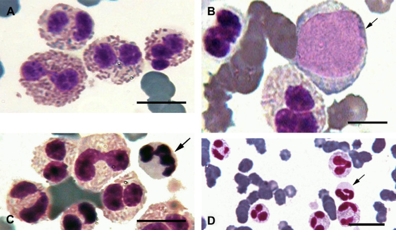
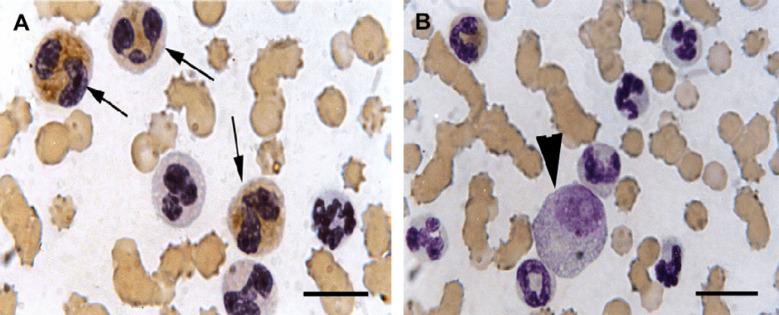
A 14-year-old female domestic shorthair cat was presented to Tehran University Veterinary Teaching Hospital for a persistent fever, anorexia, intermittent vomiting, weight loss and weakness. The main clinical signs were pale mucous membranes, dehydration and splenomegaly. The complete blood count and serum biochemistry tests revealed non-regenerative anaemia, thrombocytopenia and increased alkaline phosphatase (ALP) activity. An enzyme-linked immunosorbent assay (ELISA) test for feline leukaemia virus was negative. Blood film and bone marrow examination revealed a large number of immature eosinophils with variable sizes and numbers of faintly azurophilic granules. Cytochemical staining of blood film demonstrated 70% positive cells for ALP activity. Four percent CD34 positive cells were detected by flow cytometry. As eosinophilic leukaemia is difficult to identify by light microscopy, well-defined diagnostic criteria and the use of flow cytometry and cytochemical staining can improve the ability to correctly diagnose this type of leukaemia in cats.
Figures
References
-
- Borgfeldt C.B., Hansen B., Manthrope R. The hypereosinophilic syndrome. Report of a case with successful medical treatment following cardiac biopsy, Scandinavian Journal of Rheumatology 17, 1988, 51–54. - PubMed
-
- Brito-Babapulle F. Clonal eosinophilic disorders and the hypereosinophilic syndrome, Blood Review 11, 1997, 129–145. - PubMed
-
- Facklam N.R., Kociba G.J. Cytochemical characterization of feline leukemic cells, Veterinary Pathology 23, 1986, 155–161. - PubMed
-
- Finlay D. Eosinophilic leukaemia in the cat: a case report, Veterinary Record 116, 1985, 567. - PubMed
-
- Hendrick M. A spectrum of hypereosinophilic syndrome examplified by six cats with eosinophilic enteritis, Veterinary Pathology 18, 1981, 188–200. - PubMed
Publication types
MeSH terms
LinkOut - more resources
Full Text Sources
Research Materials
Miscellaneous
