. 2007 Oct;81(20):10861-8.
doi: 10.1128/JVI.00813-07. Epub 2007 Aug 1.

Early establishment and antigen dependence of simian immunodeficiency virus-specific CD8+ T-cell defects

Affiliations

Early establishment and antigen dependence of simian immunodeficiency virus-specific CD8+ T-cell defects

Yvonne M Mueller et al. J Virol. 2007 Oct.

Abstract

Differentiation and survival defects of human immunodeficiency virus (HIV)-specific CD8(+) T cells may contribute to the failure of HIV-specific CD8(+) T cells to control HIV replication. It is not known, however, whether simian immunodeficiency virus (SIV)-infected rhesus macaques show comparable defects in these virus-specific CD8(+) T cells or when such defects are established during infection. Peripheral blood cells from acutely and chronically infected rhesus macaques were stained ex vivo for memory subpopulations and examined by in vitro assays for apoptosis sensitivity. We show here that SIV-specific CD8(+) T cells from chronically SIV infected rhesus macaques show defects comparable to those observed in HIV infection, namely, a skewed CD45RA(-) CD62L(-) effector memory phenotype, reduced Bcl-2 levels, and increased levels of spontaneous and CD95-induced apoptosis of SIV-specific CD8(+) T cells. Longitudinal studies showed that the survival defects and phenotype are established early in the first few weeks of SIV infection. Most importantly, they appear to be antigen driven, since most probably the loss of epitope recognition due to viral escape results in the reversal of the phenotype and reduced apoptosis sensitivity, something we observed also for animals treated with antiretroviral therapy. These findings further support the use of SIV-infected rhesus macaques to investigate the phenotypic changes and apoptotic defects of HIV-specific CD8(+) T cells and indicate that such defects of HIV-specific CD8(+) T cells are the result of chronic antigen stimulation.


Figures

FIG. 1.
FIG. 1.
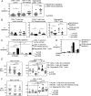
Reduced levels of CD45RA+ CD62L effector memory SIV-specific CD8+ T cells in chronically SIV infected rhesus macaques. PBMC from uninfected and SIV-infected rhesus macaques were analyzed ex vivo for memory subpopulations. Gag-specific CD8+ T cells from SIV-infected animals were identified using the Gag CM9 tetramer. (A) Representative FACS plots showing memory subpopulations of Gag-specific and total CD8+ T cells from an SIV-infected rhesus macaque after direct ex vivo staining of freshly isolated PBMC. Cells were first gated on lymphocytes using forward scatter and side scatter, then on CD3+ T cells, and subsequently for the indicated antigens. (B) Percentages of memory subpopulations in CD8+ T cells from uninfected control rhesus macaques (n = 18) and in total and Gag-specific CD8+ T cells from SIV-infected rhesus macaques (n = 15). Horizontal lines indicate means. (C) Ratios of CD45RA CD62L to CD45RA+ CD62L effector memory cells for total CD8+ T cells from uninfected animals and for Gag-specific and total CD8+ T cells from SIV-infected animals. Each line indicates a statistically significant difference between two groups. P values are given in the key.
FIG. 2.
FIG. 2.
SIV-specific CD8+ T cells are highly sensitive to apoptosis, which is inhibited by IL-15. PBMC from uninfected and SIV-infected rhesus macaques were cultured in the presence or absence of sFasL (10 ng/ml) overnight, and spontaneous and CD95-induced apoptosis was analyzed. Gag-specific CD8+ T cells from SIV-infected animals were identified using the Gag CM9 tetramer. (A) Pooled data showing the percentages of spontaneous and CD95-induced apoptosis for CD8+ T cells from uninfected rhesus macaques (n = 18) and for total (n = 15) and Gag-specific (n = 15) CD8+ T cells from SIV-infected rhesus macaques in overnight cultures of PBMC. Horizontal lines indicate means. (B) Pooled data showing the percentages of spontaneous and CD95-induced apoptosis in the presence and absence of 5 ng/ml IL-15 for CD8+ T cells from uninfected rhesus macaques (n = 18) and for total (n = 15) and Gag-specific (n = 15) CD8+ T cells from SIV-infected rhesus macaques in overnight cultures of PBMC. Horizontal lines indicate means. (C) CD95-specific apoptosis of total CD8+ T cells from uninfected rhesus macaques and of total and Gag-specific CD8+ T cells from SIV-infected rhesus macaques. For calculation of specific apoptosis, see Materials and Methods. Horizontal lines indicate means. Lines with asterisks at the top indicate statistical significance. P values are given in the key. (D) Pooled data showing the percentages of spontaneous and CD95-induced apoptosis of naïve and memory CD8+ T-cell subpopulations of chronically SIV infected rhesus macaques (n = 5). (E) (Left) Pooled data showing MFI of Bcl-2 expression in total CD8+ T cells from 16 uninfected macaques and in Gag-specific and total CD8+ T cells from 15 SIV-infected rhesus macaques. (Right) Bcl-2 MFIs after 14 h in culture in the presence or absence of IL-15 (5 ng/ml) for total CD8+ T cells from uninfected controls (n = 6) and for total (n = 7) and Gag-specific (n = 7) CD8+ T cells from SIV-infected animals. (F) (Left) MFI of Bcl-xL expression in total CD8+ T cells from 16 uninfected macaques and in Gag-specific and total CD8+ T cells from 15 SIV-infected rhesus macaques. (Right) Bcl-xL MFIs after 14 h in culture in the presence or absence of IL-15 (5 ng/ml) for total CD8+ T cells from uninfected controls (n = 6) and total (n = 7) and Gag-specific (n = 7) CD8+ T cells from SIV-infected animals.
FIG. 3.
FIG. 3.
Memory phenotype and apoptosis susceptibility of Gag-specific CD8+ T cells in acutely SIV infected rhesus macaques. (A) Pooled data for viral loads and absolute numbers of total, Gag-specific, and Tat-specific CD8+ T cells from week zero up to week 20 post-SIV infection. (B) Distribution of memory cells for total, Gag-specific, and Tat-specific CD8+ T cells from week zero up to week 20 post-SIV infection. (C) Spontaneous and CD95-induced apoptosis for total, Gag-specific, and Tat-specific CD8+ T cells from week zero up to week 20 post-SIV infection. For all panels, data for day zero to day 56 represent 12 animals, and data for day 84 to day 140 represent 5 animals. Means ± standard errors for each time point are shown.
FIG. 4.
FIG. 4.
ART treatment reverses the memory phenotype. (A) Pooled data for memory subpopulations of Gag-specific CD8+ T cells from ART-treated (n = 4) and ART-naïve (n = 11) animals. Lines indicate means. (B) Ratio of CD45RA CD62L to CD45RA+ CD62L Gag-specific CD8+ T cells from ART-treated (n = 4) and ART-naïve (n = 11) animals. Means ± standard errors are shown.

Similar articles

Cited by

References

    1. Allen, T. M., D. H. O'Connor, P. Jing, J. L. Dzuris, B. R. Mothe, T. U. Vogel, E. Dunphy, M. E. Liebl, C. Emerson, N. Wilson, K. J. Kunstman, X. Wang, D. B. Allison, A. L. Hughes, R. C. Desrosiers, J. D. Altman, S. M. Wolinsky, A. Sette, and D. I. Watkins. 2000. Tat-specific cytotoxic T lymphocytes select for SIV escape variants during resolution of primary viraemia. Nature 407:386-390. - PubMed
    1. Allen, T. M., J. Sidney, M. F. del Guercio, R. L. Glickman, G. L. Lensmeyer, D. A. Wiebe, R. DeMars, C. D. Pauza, R. P. Johnson, A. Sette, and D. I. Watkins. 1998. Characterization of the peptide binding motif of a rhesus MHC class I molecule (Mamu-A*01) that binds an immunodominant CTL epitope from simian immunodeficiency virus. J. Immunol. 160:6062-6071. - PubMed
    1. Arnoult, D., F. Petit, J. D. Lelievre, D. Lecossier, A. Hance, V. Monceaux, B. Hurtrel, R. Ho Tsong Fang, J. C. Ameisen, and J. Estaquier. 2003. Caspase-dependent and -independent T-cell death pathways in pathogenic simian immunodeficiency virus infection: relationship to disease progression. Cell Death Differ. 10:1240-1252. - PubMed
    1. Barouch, D. H., J. Kunstman, J. Glowczwskie, K. J. Kunstman, M. A. Egan, F. W. Peyerl, S. Santra, M. J. Kuroda, J. E. Schmitz, K. Beaudry, G. R. Krivulka, M. A. Lifton, D. A. Gorgone, S. M. Wolinsky, and N. L. Letvin. 2003. Viral escape from dominant simian immunodeficiency virus epitope-specific cytotoxic T lymphocytes in DNA-vaccinated rhesus monkeys. J. Virol. 77:7367-7375. - PMC - PubMed
    1. Champagne, P., G. S. Ogg, A. S. King, C. Knabenhans, K. Ellefsen, M. Nobile, V. Appay, G. P. Rizzardi, S. Fleury, M. Lipp, R. Forster, S. Rowland-Jones, R. P. Sekaly, A. J. McMichael, and G. Pantaleo. 2001. Skewed maturation of memory HIV-specific CD8 T lymphocytes. Nature 410:106-111. - PubMed

Publication types

Substances