. 2007 Oct;81(20):11208-17.
doi: 10.1128/JVI.00919-07. Epub 2007 Aug 1.

Serine/arginine-rich proteins contribute to negative regulator of splicing element-stimulated polyadenylation in rous sarcoma virus

Affiliations

Serine/arginine-rich proteins contribute to negative regulator of splicing element-stimulated polyadenylation in rous sarcoma virus

Nicole L Maciolek et al. J Virol. 2007 Oct.

Abstract

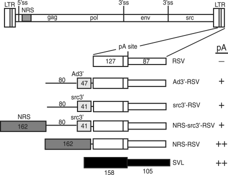
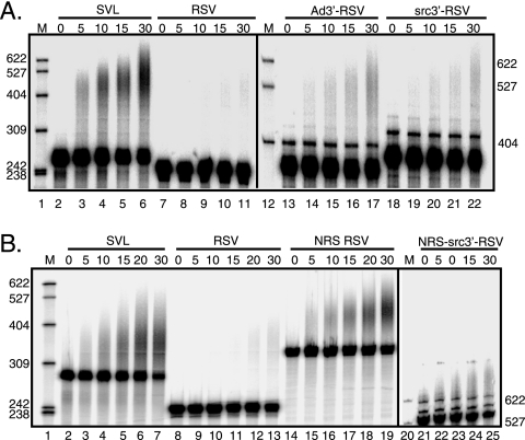
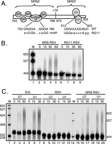
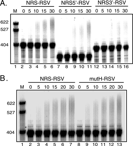
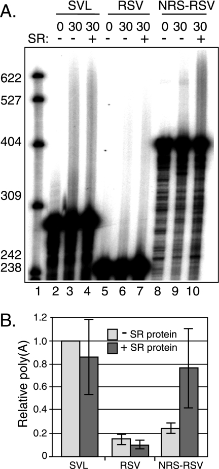
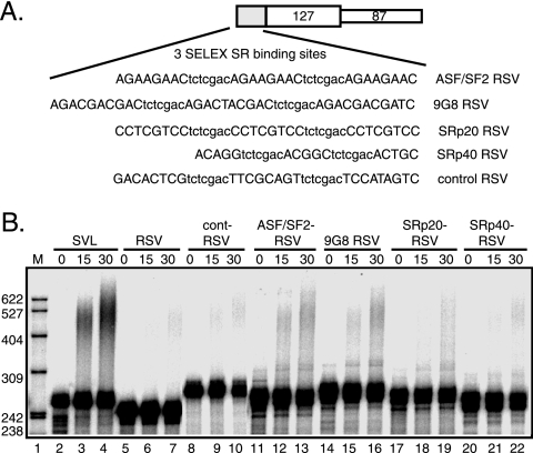
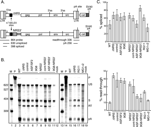
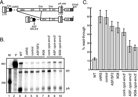
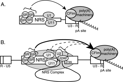
Rous sarcoma virus (RSV) requires large amounts of unspliced RNA for replication. Splicing and polyadenylation are coupled in the cells they infect, which raises the question of how viral RNA is efficiently polyadenylated in the absence of splicing. Optimal RSV polyadenylation requires a far-upstream splicing control element, the negative regulator of splicing (NRS), that binds SR proteins and U1/U11 snRNPs and functions as a pseudo-5' splice site that interacts with and sequesters 3' splice sites. We investigated a link between NRS-mediated splicing inhibition and efficient polyadenylation. In vitro, the NRS alone activated a model RSV polyadenylation substrate, and while the effect did not require the snRNP-binding sites or a downstream 3' splice site, SR proteins were sufficient to stimulate polyadenylation. Consistent with this, SELEX-binding sites for the SR proteins ASF/SF2, 9G8, and SRp20 were able to stimulate polyadenylation when placed upstream of the RSV poly(A) site. In vivo, however, the SELEX sites improved polyadenylation in proviral clones only when the NRS-3' splice site complex could form. Deletions that positioned the SR protein-binding sites closer to the poly(A) site eliminated the requirement for the NRS-3' splice site interaction. This indicates a novel role for SR proteins in promoting RSV polyadenylation in the context of the NRS-3' splice site complex, which is thought to bridge the long distance between the NRS and poly(A) site. The results further suggest a more general role for SR proteins in polyadenylation of cellular mRNAs.


Figures

FIG. 1.
FIG. 1.
Schematic of in vitro polyadenylation substrates. At the top is a diagram of the RSV provirus showing the long terminal repeats (LTR); 5′ ss; NRS (shaded); gag, pol, env, and src genes; and 3′ ss. Below is an expansion of the 3′ LTR and a schematic of the RSV substrates, which include the entire region downstream of the poly(A) site (87 nt) (thin open box) and 127 nt of upstream sequence (wide open box). Ad3′-RSV has 47 nt of Ad 3′ exon (lightly shaded box) with 80 nt of upstream intron (thin line). src3′-RSV has 41 nt of the src 3′ exon (shaded box) with 80 nt of upstream RSV intron. NRS-src3′-RSV and NRS-RSV have the 162-nt NRS BBΔ76 fragment (dark shaded box) inserted upstream of the RSV poly(A) signal, with or without the src 3′ ss region. The positive control SVL substrate (black boxes) contains 137 nt of upstream and 105 nt of downstream sequence relative to the SVL poly(A) site. A summary of polyadenylation activity is indicated at the far right (−, activity less than 10% of that of SVL; +, activity of up to 40% of that of SVL; ++, activity of 40% or more of that of SVL).
FIG. 2.
FIG. 2.
The NRS alone can stimulate RSV polyadenylation. The indicated RSV polyadenylation substrates (see Fig. 1) containing a 3′ ss (A) and/or the NRS (B) were uniformly labeled with 32P and incubated in HeLa nuclear extract for the times (in minutes) indicated above each lane. Polyadenylation appears as an upward smear. Samples were subjected to electrophoresis on a 6% 8 M urea polyacrylamide gel, and images were obtained with a PhosphorImager. Ad3′-RSV and src3′-RSV samples (A) and NRS-src3′-RSV samples (B) were run ∼1 h longer to allow adequate separation of products. The images are representative of at least three independent repeats. M, 32P-end-labeled pBR322/MspI markers, the sizes of which are indicated at the left and right. The results of quantitation of polyadenylation at 30 min are the following: (A) SVL, 32%; RSV, 2%; Ad3′-RSV, 8%; and src3′-RSV, 4%; (B) SVL, 47%; RSV, 6%; NRS-RSV, 63%; and NRS-src3′-RSV, 7%.
FIG. 3.
FIG. 3.
U1 and U11 snRNPs are not required for NRS-stimulated RSV polyadenylation. (A) Schematic of the NRS (nt 703 to 932) indicating the NRS5′- (nt 703 to 798), NRS3′- (nt 798 to 932), and NRS-binding factors. Shown are the binding of SR proteins and hnRNP H to NRS5′ and U1 and U11 snRNPs binding to NRS3′. Δ76 indicates a 76-nt deletion that does not markedly affect NRS function. The sequences of mutations that eliminate hnRNP H (mutH) and U1/U11 snRNP (RG11) binding are shown below the wild-type sequence. (B) RSV substrates harboring the wild-type NRS or the RG11 mutation were uniformly labeled with 32P, and polyadenylation was assessed in HeLa cell nuclear extract. (C) SVL, RSV, and NRS-RSV substrates were labeled as described for panel B, and polyadenylation was assayed in HeLa nuclear extract in which U1 or U7 snRNP was inactivated using 2′-O-methyl oligonucleotides. (B and C) Reaction mixtures were incubated for the times (in minutes) indicated above each lane and were subjected to electrophoresis on a 6% polyacrylamide gel that contains 8 M urea. (B) NRS-RSV samples were run ∼1 h longer to allow adequate separation of products. Polyadenylation appears as a slower-migrating smear. Images were obtained with a PhosphorImager and are representative of at least three independent experiments. M, 32P-end-labeled pBR322/MspI markers; WT, wild type. The results of quantitation of polyadenylation at 30 min are the following: (B) NRS-RSV, 9%; and RG11-RSV, 8%; (C) SVL/U7, 45%; SVL/U1, 31%; RSV/U7, 8%; RSV/U1, 5%; NRS RSV/U7, 20%; and NRS RSV/U1, 19%.
FIG. 4.
FIG. 4.
Polyadenylation stimulatory activity maps to NRS5′, but hnRNP H-binding sites are not required. (A) RNA substrates were uniformly labeled with 32P and incubated in HeLa nuclear extract for the times (in minutes) indicated above each lane. The NRS5′ and NRS3′ regions are the same as those bracketed in Fig. 3A, except that this NRS3′ version contained nt 801 to 932. RNA was subjected to electrophoresis on a 6% polyacrylamide gel that contains 8 M urea, and the image was obtained with a PhosphorImager; the results are representative of at least three independent experiments. Polyadenylation appears as a slower-migrating smear. The results of quantitation of polyadenylation at 30 min are the following: NRS-RSV, 13%; NRS5′-RSV, 12%; and NRS3′-RSV, 2%. (B) NRS-RSV and mutH-RSV, which contain the mutated hnRNP H-binding sites (Fig. 3A), were treated as described for panel A. The results of quantitation of polyadenylation at 30 min are the following: NRS-RSV, 13%; and NRSmutH-RSV, 13%. M, 32P-end-labeled pBR322/MspI markers, the sizes of which are indicated at left.
FIG. 5.
FIG. 5.
SR proteins are required for NRS-stimulated RSV polyadenylation in vitro. (A) SVL, RSV, and NRS-RSV substrates were labeled with 32P and incubated in HeLa cell S100 extract supplemented with 16% HeLa cell nuclear extract. Substrates were incubated with (+) or without (−) purified SR proteins for 30 min and were subjected to electrophoresis on a 6% polyacrylamide gel that contains 8 M urea. An image representative of three independent experiments was obtained with a PhosphorImager. Polyadenylation appears as a slower-migrating smear. M, 32P-end-labeled pBR322/MspI markers, the sizes of which are indicated at the left. (B) Quantitation of the data shown in panel A. The percent polyadenylation for SVL substrate in the absence of SR proteins was set at 1.0, and the relative levels for the RSV and NRS-RSV substrates were normalized to this value. Gray bars, without SR proteins; dark bars, with SR proteins. Error bars indicate standard deviations.
FIG. 6.
FIG. 6.
SR protein-binding sites stimulate RSV polyadenylation in vitro. (A) Schematic of RSV substrate and sequence of SR protein SELEX-binding sites inserted upstream of the RSV poly(A) site. Consensus sequences (upper case) used for ASF/SF2, 9G8, SRp20, and SRp40 are separated by a 7-nt spacer (lowercase letters) (8, 29, 51). The negative control contains three repeats of a sequence from the original ASF/SF2 SELEX pool (51). (B) SVL-, RSV-, and SELEX-containing substrates were uniformly labeled with 32P and incubated in HeLa nuclear extract for the times (in minutes) indicated above each lane. RNA was subjected to electrophoresis on a 6% polyacrylamide gel that contains 8 M urea. The image, representative of at least three independent experiments, was obtained with a PhosphorImager. Polyadenylation appears as an upward smear. M, 32P-end-labeled pBR322/MspI markers, the sizes of which are indicated at the left. The results of quantitation of polyadenylation are the following: SVL, 18%; RSV, 2%; cont-RSV, 4%; ASF/SF2-RSV, 10%; 9G8-RSV, 7%; SRp20-RSV, 9%; SRp40-RSV, 4%.
FIG. 7.
FIG. 7.
SR protein-binding sites stimulate polyadenylation of proviral clones in vivo. (A) Schematic of proviral constructs. Shown are the long terminal repeats (LTR); 5′ and 3′ ss; gag, pol, env, and src genes; poly(A) site; and downstream CAT gene and SVL poly(A) signal (shaded). At the top, the ASF/SF2 or 9G8 SELEX site (black box) replaced the entire NRS, while at the bottom, the SELEX sites were fused to NRS3′ (gray box). The positions and sizes of the RNase protection probe used for panel B and the protected products are shown below the lower schematic. (B) RNase protection assays were performed using RNA from CEFs transfected with the indicated proviral clones lacking the entire NRS (ΔNRS) or containing only NRS3′ or with constructs having insertions of control, ASF/SF2, or 9G8 SELEX sites. On the right are constructs with mutations that eliminate U1/U11 snRNP binding (RG11) or hnRNP H binding to the downstream sites (mG1+2). Products were subjected to electrophoresis on a 6% polyacrylamide gel that contains 8 M urea and visualized with a PhosphorImager. Protected products are labeled at the right. P, probe (864 nt); US, unspliced RNA (630 nt); S, spliced RNA (398 nt); RT, read-through RNA (336 nt); pA, polyadenylated product (256 nt); WT, wild type. M, 32P-end-labeled pBR322/MspI markers, the sizes of which are indicated at the left. (C) Quantitation of the data from three replicate experiments for the percentages of spliced (top) and read-through (bottom) RNA.
FIG. 8.
FIG. 8.
ASF/SF2 and 9G8 SELEX sites can activate RSV polyadenylation in vivo independently of the NRS-3′ ss complex. (A) Schematic of proviral constructs. Shown are the proviral long terminal repeats (LTRs); 5′ and 3′ ss; gag, pol, env, and src genes; poly(A) site; and downstream CAT gene and SVL poly(A) signal (shaded). The ASF/SF2 and 9G8 SELEX sites or control sequence (black box) replaced the entire NRS (ΔNRS; dashed line). The deletion (denoted by the lines) places the SELEX (or control) sequences nearer to the poly(A) site. (B) RNase protection assay (probe and products were the same as those used for Fig. 7) of total RNA isolated from CEFs transfected with the indicated proviral clones. Protected read-through and poly(A) products (designated on the right) were resolved on a 6% polyacrylamide gel that contains 8 M urea and visualized with a PhosphorImager. P, probe; RT, read-through RNA; pA, polyadenylated product; WT, wild type. M, 32P-end-labeled pBR322/MspI markers, the sizes of which are indicated at the left. Bands were quantitated using a PhosphorImager. (C) Quantitation of the three independent experiments.
FIG. 9.
FIG. 9.
Model for NRS-stimulated RSV polyadenylation in vitro and in vivo. (A) In vitro model. The NRS is shown with SR proteins and U1/U11 snRNPs bound. When in close proximity to the RSV polyadenylation site (pA site; at the end of the proviral U3-R region), as is the case with in vitro constructs, SR proteins recruit the polyadenylation machinery, possibly through an RS domain interaction with a similar domain in CFIm (arrow). (B) In vivo model. Shown are the 5′ end of viral RNA (R-U5) and the poly(A) site (pA site) at the 3′ end (U3-R). The NRS and associated factors are shown interacting with a 3′ ss to form the nonproductive NRS complex (shaded oval), which positions SR proteins at least 4,200 nt closer to the poly(A) site. SR proteins then recruit the polyadenylation machinery (arrow), possibly through CFIm. Potential interactions between the NRS-3′ ss complex factors U1, U2, and/or U2AF and the polyadenylation machinery also are shown (dotted arrows) (see the text for details). WT, wild type.

Similar articles

Cited by

References

    1. Amendt, B. A., S. B. Simpson, and C. M. Stoltzfus. 1995. Inhibition of RNA splicing at the Rous sarcoma virus src 3′ splice site is mediated by an interaction between a negative cis element and a chicken embryo fibroblast nuclear factor. J. Virol. 69:5068-5076. - PMC - PubMed
    1. Arhin, G. K., M. Boots, P. S. Bagga, C. Milcarek, and J. Wilusz. 2002. Downstream sequence elements with different affinities for the hnRNP H/H′ protein influence the processing efficiency of mammalian polyadenylation signals. Nucleic Acids Res. 30:1842-1850. - PMC - PubMed
    1. Arrigo, S., and K. Beemon. 1988. Regulation of Rous sarcoma virus RNA splicing and stability. Mol. Cell. Biol. 8:4858-4867. - PMC - PubMed
    1. Awasthi, S., and J. C. Alwine. 2003. Association of polyadenylation cleavage factor I with U1 snRNP. RNA 9:1400-1409. - PMC - PubMed
    1. Bagga, P. S., G. K. Arhin, and J. Wilusz. 1998. DSEF-1 is a member of the hnRNP H family of RNA-binding proteins and stimulates pre-mRNA cleavage and polyadenylation in vitro. Nucleic Acids Res. 26:5343-5350. - PMC - PubMed

Publication types