. 2007 Oct;81(20):10991-1004.
doi: 10.1128/JVI.00705-07. Epub 2007 Aug 1.

Herpes simplex virus type 1 genomes are associated with ND10 nuclear substructures in quiescently infected human fibroblasts

Affiliations

Herpes simplex virus type 1 genomes are associated with ND10 nuclear substructures in quiescently infected human fibroblasts

Roger D Everett et al. J Virol. 2007 Oct.

Abstract

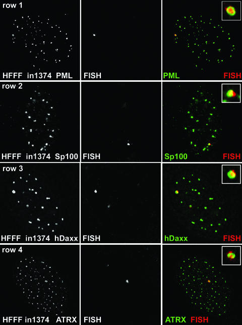
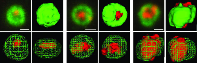
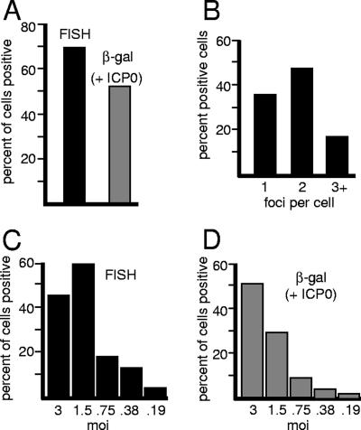
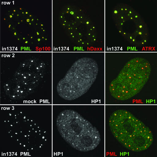
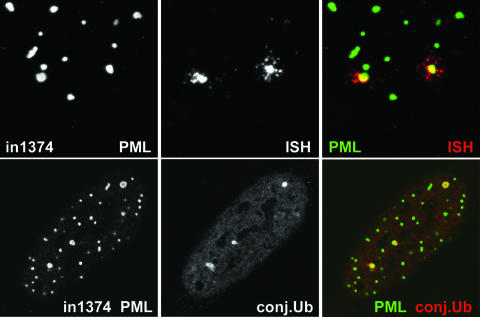
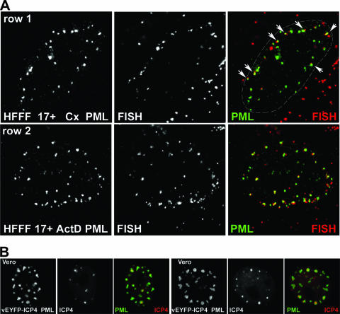
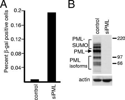
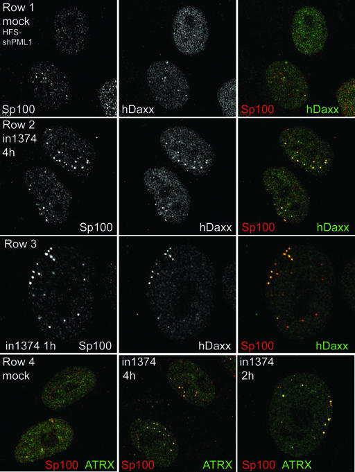
Herpes simplex virus type 1 (HSV-1) genomes become associated with structures related to cellular nuclear substructures known as ND10 or promyelocytic leukemia nuclear bodies during the early stages of lytic infection. This paper describes the relationship between HSV-1 genomes and ND10 in human fibroblasts that maintain the viral genomes in a quiescent state. We report that quiescent HSV-1 genomes detected by fluorescence in situ hybridization (FISH) are associated with enlarged ND10-like structures, frequently such that the FISH-defined viral foci are apparently enveloped within a sphere of PML and other ND10 proteins. The number of FISH viral foci in each quiescently infected cell is concordant with the input multiplicity of infection, with each structure containing no more than a small number of viral genomes. A proportion of the enlarged ND10-like foci in quiescently infected cells contain accumulations of the heterochromatin protein HP1 but not other common markers of heterochromatin such as histone H3 di- or trimethylated on lysine residue 9. Many of the virally induced enlarged ND10-like structures also contain concentrations of conjugated ubiquitin. Quiescent infections can be established in cells that are highly depleted for PML. However, during the initial stages of establishment of a quiescent infection in such cells, other ND10 proteins (Sp100, hDaxx, and ATRX) are recruited into virally induced foci that are likely to be associated with HSV-1 genomes. These observations illustrate that the intimate connections between HSV-1 genomes and ND10 that occur during lytic infection also extend to quiescent infections.


Figures

FIG. 1.
FIG. 1.
Detection of HSV-1 genomes in quiescently infected cells. HFFF cells were infected with virus in1374 (MOI, 3 PFU per cell) and then maintained at 38.5°C for 8 days before being processed for FISH detection of HSV-1 genomes (center panel of each row) and staining for ND10 components by immunofluorescence (left panels). (Row 1) PML; (row 2) Sp100; (row 3) hDaxx; (row 4) ATRX. The merged signals are shown in color on the right (proteins in green; FISH signal in red), and the insets show the FISH foci at a higher magnification.
FIG. 2.
FIG. 2.
Z-stack and 3-D reconstruction analysis of PML foci containing viral DNA. Image stacks of typical examples of viral FISH foci, as in Fig. 1, row 1, were imported into Autodeblur and Autovisualize software (Media Cybernetics) for 3-D reconstruction and production of rendered images. Three examples are shown. In each case, the top left image of the set presents a projection of the complete Z-stack onto a single plane (bar, 0.5 μm); the top right shows a 3-D reconstruction of the outer rendered surface; and the lower two images show alternative views with the PML signal (green) represented as a cage to reveal the FISH signal (red) in the interior.
FIG. 3.
FIG. 3.
Quantification of quiescently infected cells exhibiting the HSV-1-specific FISH signal. (A) HFFF cells were infected with in1374 (MOI, 3) as for Fig. 1; then, at 9 days postinfection, the samples were processed for FISH and immunofluorescence detection of PML. Four random fields of view were analyzed by counting the proportion of cells exhibiting FISH foci in association with PML. A total of 143 cells were analyzed (filled bar). At the same time, parallel samples were infected with 2 PFU of the ICP0-expressing virus tsK per cell to induce reactivation of β-galactosidase expression; then the samples were stained for β-galactosidase activity and counterstained with Carmalum. The proportion of β-galactosidase-positive cells was calculated (shaded bar). (B) The number of positive FISH foci was determined in each positive cell counted for panel A, and the proportion of positive cells exhibiting 1, 2, or 3 or more foci is shown. (C and D) The numbers of cells that exhibit FISH foci and that can be reactivated to express β-galactosidase decrease in parallel with decreasing MOIs. (C) HFFF cells were infected with in1374 at decreasing MOIs as shown, and the proportions of cells with PML-associated FISH signals were determined at 9 days postinfection as for panel A. (D) At the same time, parallel samples were infected with tsK to determine the proportions of cells from which β-galactosidase expression could be reactivated.
FIG. 4.
FIG. 4.
Recruitment of cellular proteins to PML ring structures in cells quiescently infected with in1374. HFFF cells were infected with in1374 to establish quiescently infected cells. Then, 8 days later, the samples were processed for immunofluorescence. Cells harboring quiescent viral genomes could be identified by the characteristic ring-like structures of PML. (Row 1) Cells were stained for PML (green) and Sp100, hDaxx, or ATRX (red) as indicated. (Row 2) Mock-infected HFFF cells incubated in parallel were costained for PML (red) and HP1 (green), illustrating the variable presence of HP1 within ND10 in human fibroblasts. (Row 3) HFFF cells quiescently infected with in1374 were costained for PML (red) and HP1 (green). Many of the ring-like PML structures contained prominent HP1 staining.
FIG. 5.
FIG. 5.
The HSV-1 genome foci in quiescently infected HFFF cells can have a complex nature, and the associated PML ring structures invariably contain local accumulations of conjugated ubiquitin. (Top) HFFF cells were infected with virus in1374 (MOI, 3) and then maintained at 38.5°C for 8 days before being processed for FISH detection of HSV-1 genome foci (red) and immunofluorescence staining for PML (green). Images show details of a cell with prominent FISH foci that exhibit a complex nature, with smaller foci surrounding the main central portion. (Bottom) HFFF cells quiescently infected with in1374 were costained for PML (green) and conjugated ubiquitin (conj.Ub) (red). As in Fig. 4, the quiescently infected cells could be identified by the presence of two or more ring-like PML structures. Such rings invariably contained localized concentrations of conjugated ubiquitin.
FIG. 6.
FIG. 6.
(A) HSV-1 gene expression is not required for the recruitment of ND10 proteins to the sites of incoming viral genomes. HFFF cells were infected with wt HSV-1 17+ (MOI, 10). At 4 h after infection, the cells were processed for FISH detection of viral genomes (red) and immunofluorescence detection of PML (green) as indicated. For rows 1 and 2, the cells had been pretreated for 15 min with cycloheximide (Cx) (100 μg/ml) or actinomycin D (ActD) (2.5 μg/ml), respectively, before the addition of virus; then infections were conducted, and the cells were maintained in the presence of the drugs throughout. At the early times of a high-MOI infection, many viral particles remain bound to the cell surface or are present in the cytoplasm, giving the extranuclear punctate red staining seen in these images. Prominent examples of genomes associated with PML are arrowed in the merged image of row 1, and the nuclear periphery is delineated by a dashed line. The genome foci at the lower right of this image are from particles that have remained adsorbed to the cell surface or in the cytoplasm. (B) Recruitment of exogenously expressed PML to ring-like foci that surround accumulations of ICP4 during the early times of a low-multiplicity infection. Vero cells were infected with a baculovirus that expresses ECFP-linked PML from the HCMV promoter. On the following day, the cells were infected with HSV-1 vEYFP-ICP4 at an MOI of 0.01. The cells were monitored by live-cell microscopy. Two cells exhibiting ICP4 foci enveloped by PML are shown. The set of images on the left shows a cell at 150 min postinfection, and that on the right shows a cell after 4 h, in both cases prior to the onset of DNA replication and before ICP0 had been able to degrade the highly overexpressed PML (approximately 100-fold greater than the level of endogenous PML [16]).
FIG. 7.
FIG. 7.
A proportion of PML-depleted human fibroblasts have a reduced ability to repress β-galactosidase expression from in1374. (A) HFS cells transduced with lentiviruses expressing an anti-luciferase control shRNA or an anti-PML shRNA were infected with in1374 (MOI, 3). On the following day, the cells were stained for β-galactosidase expression, and the number of blue cells in wells containing approximately 1 × 10⁵ cells in total was determined. A parallel control coinfection of samples of the same cells with in1374 and tsK indicated that in both cases more than 50% of the cells had received in1374 genomes (data not shown). The increases in blue cell numbers in PML-depleted cells over those in control cells in a series of repeated experiments were as follows: 17.0-, 31.0-, 32.5-, 22.7-, 12.8-, 30.1-, 19.3-, and 19.2-fold. (B) Western blot illustrating PML expression in typical isolates of fibroblasts transduced with a lentivirus expressing a control anti-luciferase shRNA or an anti-PML shRNA.
FIG. 8.
FIG. 8.
Initial recruitment of ND10 proteins Sp100, hDaxx, and ATRX into in1374-induced foci in PML-depleted cells. Human fibroblasts transduced with a lentivirus expressing an anti-PML shRNA (HFS-shPML1 cells) were infected with in1374 (MOI, 10) and then fixed at 1, 2, 4, and 6 h after infection. Mock-infected cells were fixed in parallel. Coverslips were stained with combinations of anti-Sp100 (rat sp26) and rabbit anti-hDaxx or rabbit anti-ATRX antibodies as indicated. (Row 1) Mock-infected HFS-shPML1 cells exhibit variable foci of Sp100 and mainly diffuse hDaxx staining, with little colocalization. (Row 2) HFS-shPML1 cells infected with in1374 contain pronounced foci of colocalizing Sp100 and hDaxx. (Row 3) This effect was visible in some cells as early as 1 h after virus adsorption. (Row 4) There is little colocalization between Sp100 and ATRX in mock-infected HFS-shPML1 cells (left), but pronounced colocalizing foci form at early times of in1374 infection (center and right).

Similar articles

Cited by

References

    1. Ace, C. I., T. A. McKee, J. M. Ryan, J. M. Cameron, and C. M. Preston. 1989. Construction and characterization of a herpes simplex virus type 1 mutant unable to transinduce immediate-early gene expression. J. Virol. 63:2260-2269. - PMC - PubMed
    1. Antón, L. C., U. Schubert, I. Bacik, M. F. Princiotta, P. A. Wearsch, J. Gibbs, P. M. Day, C. Realini, M. C. Rechsteiner, J. R. Bennink, and J. W. Yewdell. 1999. Intracellular localization of proteasomal degradation of a viral antigen. J. Cell Biol. 146:113-124. - PMC - PubMed
    1. Boddy, M. N., K. Howe, L. D. Etkin, E. Solomon, and P. S. Freemont. 1996. PIC 1, a novel ubiquitin-like protein which interacts with the PML component of a multiprotein complex that is disrupted in acute promyelocytic leukaemia. Oncogene 13:971-982. - PubMed
    1. Cantrell, S. R., and W. A. Bresnahan. 2006. Human cytomegalovirus (HCMV) UL82 gene product (pp71) relieves hDaxx-mediated repression of HCMV replication. J. Virol. 80:6188-6191. - PMC - PubMed
    1. Davison, M. J., V. G. Preston, and D. J. McGeoch. 1984. Determination of the sequence alteration in the DNA of the herpes simplex virus type 1 temperature-sensitive mutant ts K. J. Gen. Virol. 65:859-863. - PubMed

Publication types

Substances

LinkOut - more resources