. 2007 Oct;81(20):10981-90.
doi: 10.1128/JVI.01033-07. Epub 2007 Aug 1.

The coronavirus spike protein induces endoplasmic reticulum stress and upregulation of intracellular chemokine mRNA concentrations

Affiliations

The coronavirus spike protein induces endoplasmic reticulum stress and upregulation of intracellular chemokine mRNA concentrations

Gijs A Versteeg et al. J Virol. 2007 Oct.

Abstract

Murine hepatitis virus (MHV) and severe acute respiratory syndrome (SARS) coronavirus (CoV) are two of the best-studied representatives of the family Coronaviridae. During CoV infection, numerous cytokines and chemokines are induced in vitro and in vivo. Human interleukin 8 and its mouse functional counterpart, CXCL2, are early-expressed chemokines. Here we show that SARS-CoV and MHV induce endoplasmic reticulum (ER) stress and Cxcl2 mRNA transcription during infection in vitro. Expression of the viral spike protein significantly induced ER stress and Cxcl2 mRNA upregulation, while expression of the other structural genes did not. Additional experiments with UV-inactivated virus, cell-cell fusion-blocking antibodies, and an MHV mutant with a defect in spike protein maturation demonstrated that spike-host interactions in the ER are responsible for the induction of ER stress and subsequent Cxcl2 mRNA transcription. Despite significant increases in levels of Cxcl2 mRNA and functional nucleus-to-cytoplasm RNA transport, no CXCL2 protein was released into the medium from MHV-infected cells. Yet Sendai virus-infected cells showed substantial Cxcl2 mRNA induction and a simultaneous increase in levels of secreted CXCL2 protein. Our results demonstrate that expression of CoV spike proteins induces ER stress, which could subsequently trigger innate immune responses. However, at that point in infection, translation of host mRNA is already severely reduced in infected cells, preventing the synthesis of CXCL2 and ER stress proteins despite their increased mRNA concentrations.


Figures
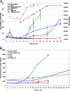
FIG. 1.
FIG. 1.
MHV and SARS-CoV induce Cxcl2 mRNA expression but prevent CXCL2 protein synthesis. L cells were infected with MHV, or L-ACE2 cells were infected with SARS-CoV, at an MOI of 10. (A) Release of infectious MHV was determined by a plaque assay, and isolated total RNA was analyzed for Cxcl2 expression by RT-qPCR. Values were normalized to GAPDH and expressed as induction (n-fold). RU, relative units. (B) Supernatants from MHV-infected L cells and SARS-CoV-infected L-ACE2 cells were analyzed for protein production by a CXCL2-specific ELISA. SeV infection was used as a positive control.
FIG. 2.
FIG. 2.
MHV spike protein induces Cxcl2 mRNA and protein expression. MHV structural genes were transiently expressed in L cells by using a vaccinia virus T7 polymerase system. (A) Cxcl2 mRNA concentrations were determined by RT-qPCR, and CXCL2 protein concentrations in the supernatant were analyzed by ELISA. RU, relative units. (B) Proteins were metabolically labeled, and expression of viral proteins was determined by immunoprecipitation with MHV-specific antibodies and analysis by SDS-polyacrylamide gel electrophoresis. Ctrl, control.
FIG. 3.
FIG. 3.
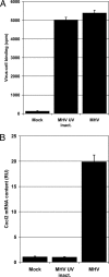
MHV-cell binding is not required for Cxcl2 induction. L cells were incubated on ice with radioactively labeled MHV or an equivalent amount of UV-inactivated (inact.) MHV. (A) After extensive removal of unbound virus, radioactivity was determined as a measure of virus binding. (B) Parallel samples were further incubated at 37°C. At 8.5 h p.i., total RNA was harvested and Cxcl2 mRNA concentrations were determined by RT-qPCR. RU, relative units.
FIG. 4.
FIG. 4.
Syncytium formation and progeny virus-cell binding are not involved in Cxcl2 induction during MHV infection. L cells were infected with MHV. At 3 h p.i., virus binding and syncytium formation were blocked by addition of neutralizing antibodies (Ab) or a control serum from nonimmunized rabbits to the medium. At 8.5 h p.i., the Cxcl2 mRNA concentration was determined by RT-qPCR (A) and cell-cell fusion was assessed by MHV-specific immunofluorescence microscopy (B). RU, relative units.
FIG. 5.
FIG. 5.
Cxcl2 induction during MHV infection is initiated by spike interactions in the ER. L cells were infected at the permissive temperature with wild-type (wt) MHV or ts379, the spike protein of which is retained in the ER at the restrictive temperature. (A) After incubation for 16 h at the permissive (34°C) or 8.5 h at the restrictive (39.5°C) temperature, Cxcl2 concentrations were determined by RT-qPCR. RU, relative units. (B) Lack of cell-cell fusion as a control for abolished spike maturation during ts379 infection at the restrictive temperature was confirmed by MHV-specific immunofluorescence microscopy.
FIG. 6.
FIG. 6.
MHV infection and SARS-CoV infection induce ER stress. (A) L cells were infected with MHV, or L-ACE2 cells were infected with SARS-CoV, at an MOI of 10. Total RNA was analyzed for Herpud1 expression by RT-qPCR. Values were GAPDH normalized and expressed as induction (n-fold). RU, relative units. (B) The same samples were analyzed for Xbp1 mRNA splicing during MHV (top) or SARS-CoV (bottom) infection by specific PCR and 2% agarose gel electrophoresis.
FIG. 7.
FIG. 7.
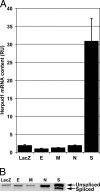
MHV spike protein induces Herpud1 mRNA. MHV structural genes were transiently expressed in L cells using a vaccinia virus T7 polymerase system. (A) Herpud1 mRNA concentrations were determined by RT-qPCR. RU, relative units. (B) The same samples were analyzed for Xbp1 mRNA splicing by specific PCR and 2% agarose gel electrophoresis.
FIG. 8.
FIG. 8.
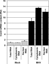
The absence of protein synthesis does not result from disturbed mRNA export from the nucleus. L cells were infected with MHV at an MOI of 10. Total, cytoplasmic, and nuclear RNAs were isolated at 7 h p.i. and analyzed for Cxcl2 expression by RT-qPCR. Values were GAPDH normalized and expressed as induction (n-fold). RU, relative units.

Similar articles

Cited by

References

    1. Banerjee, S., K. Narayanan, T. Mizutani, and S. Makino. 2002. Murine coronavirus replication-induced p38 mitogen-activated protein kinase activation promotes interleukin-6 production and virus replication in cultured cells. J. Virol. 76:5937-5948. - PMC - PubMed
    1. Bensaude, E., J. L. Turner, P. R. Wakeley, D. A. Sweetman, C. Pardieu, T. W. Drew, T. Wileman, and P. P. Powell. 2004. Classical swine fever virus induces proinflammatory cytokines and tissue factor expression and inhibits apoptosis and interferon synthesis during the establishment of long-term infection of porcine vascular endothelial cells. J. Gen. Virol. 85:1029-1037. - PubMed
    1. Bos, E. C., L. Heijnen, W. Luytjes, and W. J. Spaan. 1995. Mutational analysis of the murine coronavirus spike protein: effect on cell-to-cell fusion. Virology 214:453-463. - PMC - PubMed
    1. Bos, E. C., W. Luytjes, and W. J. Spaan. 1997. The function of the spike protein of mouse hepatitis virus strain A59 can be studied on virus-like particles: cleavage is not required for infectivity. J. Virol. 71:9427-9433. - PMC - PubMed
    1. Bos, E. C., W. Luytjes, H. V. van der Meulen, H. K. Koerten, and W. J. Spaan. 1996. The production of recombinant infectious DI-particles of a murine coronavirus in the absence of helper virus. Virology 218:52-60. - PMC - PubMed

Publication types

MeSH terms

LinkOut - more resources