Comparative Study
. 2007 Aug 1;27(31):8202-18.
doi: 10.1523/JNEUROSCI.2199-07.2007.

Chronic nicotine cell specifically upregulates functional alpha 4* nicotinic receptors: basis for both tolerance in midbrain and enhanced long-term potentiation in perforant path

Affiliations
Comparative Study

Chronic nicotine cell specifically upregulates functional alpha 4* nicotinic receptors: basis for both tolerance in midbrain and enhanced long-term potentiation in perforant path

Raad Nashmi et al. J Neurosci. .

Abstract

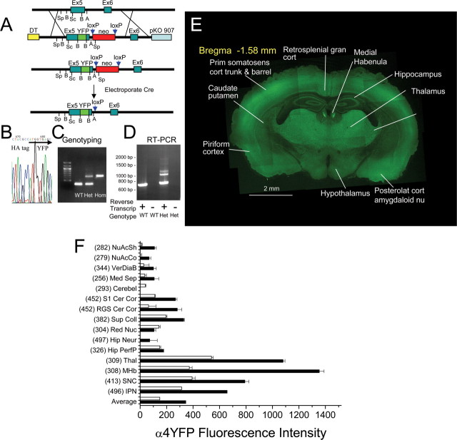
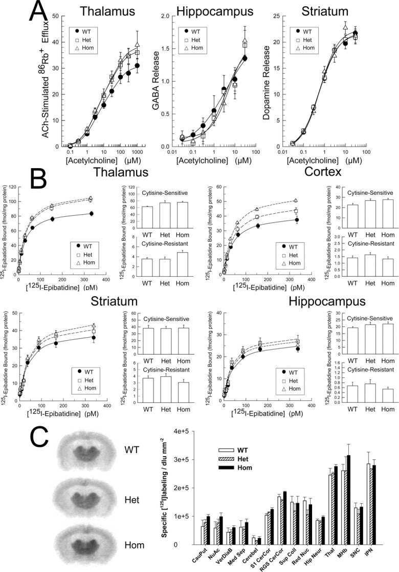
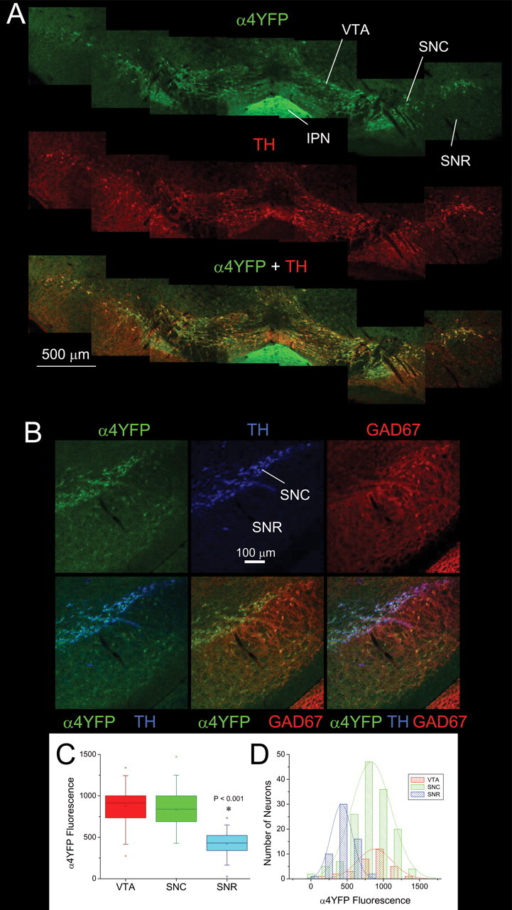
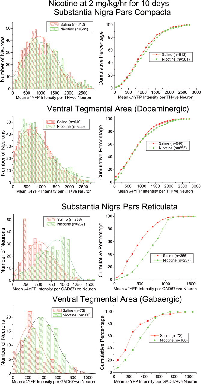
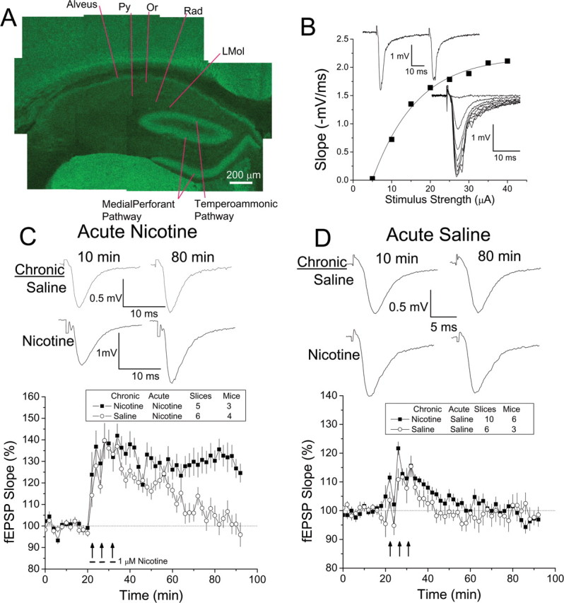
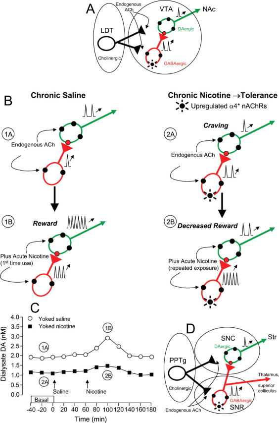
Understanding effects of chronic nicotine requires identifying the neurons and synapses whose responses to nicotine itself, and to endogenous acetylcholine, are altered by continued exposure to the drug. To address this problem, we developed mice whose alpha4 nicotinic receptor subunits are replaced by normally functioning fluorescently tagged subunits, providing quantitative studies of receptor regulation at micrometer resolution. Chronic nicotine increased alpha4 fluorescence in several regions; among these, midbrain and hippocampus were assessed functionally. Although the midbrain dopaminergic system dominates reward pathways, chronic nicotine does not change alpha4* receptor levels in dopaminergic neurons of ventral tegmental area (VTA) or substantia nigra pars compacta. Instead, upregulated, functional alpha4* receptors localize to the GABAergic neurons of the VTA and substantia nigra pars reticulata. In consequence, GABAergic neurons from chronically nicotine-treated mice have a higher basal firing rate and respond more strongly to nicotine; because of the resulting increased inhibition, dopaminergic neurons have lower basal firing and decreased response to nicotine. In hippocampus, chronic exposure to nicotine also increases alpha4* fluorescence on glutamatergic axons of the medial perforant path. In hippocampal slices from chronically treated animals, acute exposure to nicotine during tetanic stimuli enhances induction of long-term potentiation in the medial perforant path, showing that the upregulated alpha4* receptors in this pathway are also functional. The pattern of cell-specific upregulation of functional alpha4* receptors therefore provides a possible explanation for two effects of chronic nicotine: sensitization of synaptic transmission in forebrain and tolerance of dopaminergic neuron firing in midbrain.


Figures

Figure 1.
Figure 1.
Construction of the α4YFP mouse and whole-brain fluorescence. A, Genomic map of the wild-type mouse α4 nAChR allele located on chromosome 2 (top). Targeting vector was constructed with the α4 gene including exons 5 and 6 and the YFP mutation inserted in the pKO V907 vector (second line; note an inserted HA-epitope tag immediately upstream of YFP is not shown). The neomycin-resistance gene (neo) flanked by loxP sites and the diphtheria toxin A (DT) gene were also inserted as positive and negative selection markers. Map of the recombined α4 gene (third line). The neo cassette was deleted by electroporating the neo-intact ES cells with a cytomegalovirus-Cre plasmid to generate neo-deleted ES cell lines (fourth line). Restriction sites: A, AvrII; B, BstEII; Sc, SacI; Sp, SpeI. B, Chromatogram showing the start of the YFP sequence and the upstream HA-epitope tag in the α4 nAChR gene. C, PCR genotyping of WT, Het, and Hom α4YFP mice. D, RT-PCR showing α4 mRNA expression from combined thalamic–ventral midbrain tissue from Het and WT mice. The higher molecular weight band in the Het is message from the α4YFP allele. Negative controls, lacking reverse transcriptase in the RT-PCR, were devoid of bands. E, Montage of tiled confocal images of a coronal brain section at the hippocampal level from a Hom α4YFP mouse showing overall brain distribution of α4YFP. F, Quantification of α4YFP fluorescence using spectral confocal imaging taken from various brain regions from brain sections of WT, Het, and Hom α4YFP mice. The horizontal bar plots show mean α4YFP intensity (±SEM) for Het and Hom mice for each brain region. Residual autofluorescence intensity from WT mice are shown in brackets and subtracted from the raw intensity values from Het and Hom. CauPut, Caudate–putamen; NuAcSh, nucleus accumbens shell; NuAcCo, nucleus accumbens core; VerDiaB, vertical diagonal band; Med Sep, medial septum; Cerebel, cerebellar cortex; S1 CerCort, primary somatosensory cortex of barrel and trunk; RGS Cer Cor, retrosplenial granular cerebral cortex; Sup Coll, superior colliculus; Red Nuc, red nucleus; Hip Neur, hippocampal interneurons; Hip PerfP, hippocampal medial perforant path; Thal, dorsolateral geniculate nucleus of thalamus; MHb, medial habenula; IPN, interpeduncular nucleus. The “Average” bars show the α4YFP fluorescence averaged for the various regions for both Het and Hom mice.
Figure 2.
Figure 2.
Ion flux, transmitter release, agonist binding, and immunohistochemistry data. A, Left, Dose–response relationships for ACh evoked 86Rb+ efflux measurements from synaptosomes made from thalamic tissue from WT, Het, and Hom α4YFP mice. Data were fitted to two (H, L) components. For WT, EC50H = 2.6 ± 0.7 μm, EC50L = 79 ± 35 μm; Het, EC50H = 3.2 ± 0.4 μm, EC50L = 54 ± 9 μm; Hom, EC50H = 2.8 ± 2.7 μm, EC50L = 75 ± 110 μm. Middle, Right: Dose–response relationships of GABA (middle) and dopamine (right) release from the hippocampal and striatal synaptosomes, respectively, from WT, Het, and Hom α4YFP mice. Data were fitted to a single component including a Hill coefficient (n). GABA release: WT, EC50 = 3.7 ± 0.49 μm, n = 0.56 ± 0.25; Het, EC50 = 3.5 ± 1.34 μm, n = 0.94 ± 0.29; Hom, EC50 = 5.1 ± 1.8 μm, n = 1.0 ± 0.32. Dopamine release: WT, EC50 = 0.57 ± 0.09 μm, n = 0.93 ± 0.14; Het, EC50 = 0.56 ± 0.05 μm, n = 0.97 ± 0.09; Hom, EC50 = 0.61 ± 0.18 μm, n = 1.0 ± 0.29. B, Concentration dependence of 125I-epibatidine binding in the membrane fraction of thalamus, cortex, striatum, and hippocampus from WT, Het, and Hom α4YFP mice. 125I-Epibatidine binding was performed in the presence and absence of cytisine to determine the cytisine-sensitive and cytisine-resistant sites. The cytisine-sensitive binding sites are likely α4β2 nAChRs. C, Quantitative autoradiography of 125I-mAb 299 labeling in WT, Het, and Hom α4YFP mice. Representative coronal sections are shown at the hippocampal level. Nonspecific labeling was defined in parallel sets of sections from α4−/− mice, and was subtracted from total signal to define regional specific labeling. The histogram shows mean regional specific labeling densities (±SD) for each genotype. Abbreviations are as in Figure 1H.
Figure 3.
Figure 3.
Staining and quantification of dopaminergic and GABAergic α4YFP-containing neurons in the SN and VTA. A, Double labeling showing α4YFP (green) in most TH+ dopaminergic (red) neurons in the VTA and SNC. Intense α4YFP labeling is found in the interpeduncular nucleus (IPN) and light α4YFP fluorescence is found in the SNR. B, Triple labeling at higher magnification showing strong α4YFP (green) fluorescence in dopaminergic neurons (TH; blue) of the SNC and moderate α4YFP fluorescence in GABAergic neurons (GAD67; red) of the SNR. C, D, Quantification of α4YFP intensities per TH+ dopaminergic and GAD67+ GABAergic neuron in either the VTA, SNC, or SNR are shown with box plots (C) and histogram distributions (D).
Figure 4.
Figure 4.
Histograms and cumulative plots showing changes in α4YFP fluorescence in dopaminergic and GABAergic neurons of the SN and VTA with chronic nicotine. Histogram and cumulative plots of the mean α4YFP fluorescence per TH+ (dopaminergic) or GAD67+ (GABAergic) neuron from α4YFP Hom mice treated either with chronic saline or chronic nicotine (2 mg · kg−1 · h−1) for 10 d. α4YFP fluorescence was imaged from VTA, SNC, and SNR from brain sections triple labeled with TH, GAD67, and α4YFP. Dopaminergic neurons in SNC and VTA show very little change in α4YFP. However, GABAergic neurons in VTA and SNR show a shift (increase) in α4YFP intensity with chronic nicotine.
Figure 5.
Figure 5.
Patch recordings from midbrain GABAergic neurons in slices from mice exposed to chronic nicotine or saline. A, Chronic nicotine treatment enhanced baseline firing rate in SNR GABAergic neurons. Typical traces (0.5 s) (a, baseline; b, peak; c, wash) and time course of firing also show that puff application (5 s) of nicotine (1 μm) increased firing rate in neurons from chronic saline (top traces and empty circles)- and nicotine (bottom traces and filled circles)-treated mice. B, C, Summary of nicotine-induced increment of firing rate in SNR GABAergic neurons. Data in time course and summary are shown as mean ± SEM.
Figure 6.
Figure 6.
Patch recordings from midbrain dopaminergic neurons in slices from mice exposed to chronic nicotine or saline. A, Chronic nicotine treatment decreased the baseline firing rate of SNC dopaminergic neurons. Typical traces (3 s) and time course of firing also show that nicotine (1 μm) bath perfusion increased firing rate in SNC dopaminergic neurons from chronic saline (empty circles)- and nicotine (filled circles) (a, baseline; b, peak; c, wash)-treated mice. B, C, Summary of nicotine-induced increment of firing rate in SNC dopaminergic neurons. Data in time course and summary are shown as mean ± SEM.
Figure 7.
Figure 7.
α4YFP in the hippocampus and LTP experiments on the medial perforant path. A, A montage of tiled spectral confocal images of the hippocampus from a Hom α4YFP mouse, showing strong α4YFP fluorescence in axonal fiber tracts including the medial perforant path, the temperoammonic path, and the alveus. Py, Pyramidal cell layer; Or, stratum oriens; Rad, stratum radiatum; L Mol, stratum lacunosum molecular. B, An input–output relationship for fEPSP response slopes. The bottom inset shows the traces with varying stimulus intensities. The stimulus intensity eliciting 40% of the maximal slope was used for baseline stimulus and LTP induction. The top inset shows a paired-pulse stimulation with 50 ms interstimulus interval. The paired-pulse inhibition confirms that stimulation and recording were done in the medial perforant path. C, Traces and plots are shown for fEPSP slope responses on LTP induction recorded from the medial perforant path in hippocampal slices from mice chronically treated with saline or nicotine (2 mg · kg−1 · h−1 for 10–14 d). Note that fEPSP waveforms are an average of 10 traces. Slices receiving acute nicotine (1 μm) and tetanic stimuli in chronic nicotine-treated mice had larger LTP induction than chronic saline-treated mice. D, Traces and plots of fEPSP slope responses in medial perforant path in hippocampal slices from mice receiving either chronic saline or chronic nicotine. With tetanic stimuli and acute saline, there was no induction of LTP from slices in both chronic saline- and chronic nicotine-treated mice.
Figure 8.
Figure 8.
Possible consequences of upregulated functional α4 receptors on midbrain GABAergic neurons. A, Simplified circuit diagram showing the cholinergic lateral tegmental nucleus (LDT) projecting to VTA, in which ∼10% of neurons are GABAergic neurons and inhibit the dopaminergic neurons. Both GABAergic and dopaminergic neurons express α4* receptors. The VTA dopaminergic neurons project mainly to the nucleus accumbens. B, C, Events in the nicotine-naive and upregulated VTA, explaining contemporary data from animals yoked to other animals that self-administer nicotine (C) (Rahman et al., 2004). In a nicotine-naive animal, endogenous ACh modestly excites GABAergic neurons; and first exposure to nicotine robustly activates dopaminergic neurons (“Yoked saline” in C). After chronic nicotine (“Yoked nicotine” in C), endogenous ACh provides increased inhibitory tone caused by increased α4* receptors in GABAergic neurons. This leads to decreased basal dopamine release, as measured in C. Then, a nicotine dose activates the GABAergic neurons more than previously, blunting activation of the dopaminergic neurons and dopamine release, as measured in C. D, The pedunculopontine tegmental nucleus (PPTg) provides the major cholinergic projection to SN. In SN, the GABAergic and dopaminergic neurons are mostly segregated into SNR and SNC, respectively. As in VTA, both cell types have α4* receptors. SNR neurons have important inhibitory projections to SNC (Mailly et al., 2003) as well as to thalamus and other regions. The events pictured in B and C would also apply to SNC.

Similar articles

Cited by

References

    1. Alkondon M, Albuquerque EX. Nicotinic receptor subtypes in rat hippocampal slices are differentially sensitive to desensitization and early in vivo functional up-regulation by nicotine and to block by bupropion. J Pharmacol Exp Ther. 2005;313:740–750. - PubMed
    1. Azam L, Chen Y, Leslie FM. Developmental regulation of nicotinic acetylcholine receptors within midbrain dopamine neurons. Neuroscience. 2007;144:1347–1360. - PMC - PubMed
    1. Bliss TV, Lomo T. Long-lasting potentiation of synaptic transmission in the dentate area of the anaesthetized rabbit following stimulation of the perforant path. J Physiol (Lond) 1973;232:331–356. - PMC - PubMed
    1. Brunzell DH, Russell DS, Picciotto MR. In vivo nicotine treatment regulates mesocorticolimbic CREB and ERK signaling in C57BL/6J mice. J Neurochem. 2003;84:1431–1441. - PubMed
    1. Cadoni C, Di Chiara G. Differential changes in accumbens shell and core dopamine in behavioral sensitization to nicotine. Eur J Pharmacol. 2000;387:R23–R25. - PubMed

Publication types

MeSH terms

LinkOut - more resources