Comparative Study
. 2007 Aug 1;27(31):8226-37.
doi: 10.1523/JNEUROSCI.5126-06.2007.

Control of chronic pain by the ubiquitin proteasome system in the spinal cord

Affiliations
Comparative Study

Control of chronic pain by the ubiquitin proteasome system in the spinal cord

Michael H Ossipov et al. J Neurosci. .

Abstract

Chronic pain is maintained in part by long-lasting neuroplastic changes in synapses and several proteins critical for synaptic plasticity are degraded by the ubiquitin-proteasome system (UPS). Here, we show that proteasome inhibitors administered intrathecally or subcutaneously prevented the development and reversed nerve injury-induced pain behavior. They also blocked pathological pain induced by sustained administration of morphine or spinal injection of dynorphin A, an endogenous mediator of chronic pain. Proteasome inhibitors blocked mechanical allodynia and thermal hyperalgesia in all three pain models although they did not modify responses to mechanical stimuli, but partially inhibited responses to thermal stimuli in control rats. In the spinal cord, these compounds abolished the enhanced capsaicin-evoked calcitonin gene-related peptide (CGRP) release and dynorphin A upregulation, both elicited by nerve injury. Model experiments demonstrated that the inhibitors may act directly on dynorphin-producing cells, blocking dynorphin secretion. Thus, the effects of proteasome inhibitors on chronic pain were apparently mediated through several cellular mechanisms indispensable for chronic pain, including those of dynorphin A release and postsynaptic actions, and of CGRP secretion. Levels of several UPS proteins were reduced in animals with neuropathic pain, suggesting that UPS downregulation, like effects of proteasome inhibitors, counteracts the development of chronic pain. The inhibitors did not produce marked or disabling motor disturbances at doses that were used to modify chronic pain. These results suggest that the UPS is a critical intracellular regulator of pathological pain, and that UPS-mediated protein degradation is required for maintenance of chronic pain and nociceptive, but not non-nociceptive responses in normal animals.


Figures
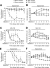
Figure 1.
Figure 1.
The proteasome inhibitors epoxomicin or MG132 prevent behavioral signs of neuropathic pain in rats with SNL. A–F, Responsive thresholds to innocuous mechanical (von Frey filaments; A, C, E) and thermal (52°C hot-plate) stimuli (B, D, F). The rats received twice-daily spinal injections of either epoxomicin at a high (0.6 nmol; A, B) or low dose (0.3 nmol; E, F) or vehicle, or received systemic injections of MG132 (5 mg/kg per injection, s.c.; C, D) beginning on the day of sham or SNL surgery. The top bars indicate the time periods of drug administration. Responses of proteasome-treated animals with SNL were significantly greater than those of vehicle-treated animals with SNL (two-factor ANOVA). SNL produced significant reductions in response parameters in vehicle-treated animals, and after termination of administration of the proteasome inhibitors. Error bars indicate mean ± SEM. n = 6–7 per group.
Figure 2.
Figure 2.
The proteasome inhibitors epoxomicin and MG132 reverse behavioral neuropathic pain in rats with SNL. A–D, Responsive thresholds to innocuous mechanical (von Frey filaments; A, C) and thermal (52°C hot-plate; B, D) stimuli. SNL produced significant reductions in paw withdrawal thresholds and hot-plate latencies 8 d after surgery (SNL; ANOVA). Rats received twice-daily subcutaneous injections of either spinal epoxomicin (0.6 nmol per injection) or systemic MG132 (5 mg/kg/injection) or vehicle starting on day 9 and finishing on day 13. Paw withdrawal thresholds and hot-plate latencies were monitored daily before the first injection of the day. Treatment with proteasome inhibitors produced significant reversals in the enhanced behavioral responses (two-factor ANOVA) that returned to baseline values after termination of administration of the proteasome inhibitor. The top bars indicate time periods of inhibitor administration. Error bars indicate mean ± SEM. n = 4–6 per group.
Figure 3.
Figure 3.
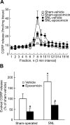
Epoxomicin normalizes evoked CGRP release after SNL. The dorsal quadrants ipsilateral to the side of injury in the spinal cord of rats treated for 7 d with vehicle or epoxomicin (twice-daily i.t. injections of 0.6 nmol) were taken 18 h after the last injection, minced, and superfused with Kreb's buffer. Epoxomicin or vehicle injections were started at the same time as the SNL or sham surgeries. A, Release was induced by capsaicin (1 μm; infusion from 12 to 18 min) and CGRP content was determined in 3 min superfusate fractions and presented as femtomoles of tissue per milligram fraction. A delay in the peak of CGRP release after capsaicin activation was caused by transit time in the tubing. B, Epoxomicin effects on capsaicin-induced CGRP release (peptide levels above baseline) after sham operation or SNL. Capsaicin-induced release in sham-operated, vehicle-treated animals was taken as 100%. Error bars indicate mean ± SEM of the percentage of induced release obtained from sham-operated rats. *,#p ≤ 0.05, comparison between sham- and SNL-operated animals injected with vehicle (*) or between respective vehicle and epoxomicin groups (#); n = 7 per group.
Figure 4.
Figure 4.
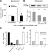
Effects of proteasome inhibitors on the PDYN system. A, Epoxomicin blocks upregulation of dynorphin A in the spinal cord of SNL rats. The dorsal quadrants ipsilateral to the side of injury in the spinal cord of rats treated for 7 d with epoxomicin (twice-daily i.t. injections of 0.6 nmol) were analyzed. B–D, Effects of epoxomicin on the PDYN levels in mouse insulinoma MIN6 cells and the dynorphin A secretion by these cells into the medium. B, PDYN was analyzed in cell extracts by Western blotting. C, D, Dynorphin A levels were measured in the culture medium by RIA. B, C, Cells were grown for 16 h in the presence of 20 or 200 nm epoxomicin, 100 nm MG132, 5 μm clasto-lactacystine β-lactone, 1 μm ALLM, or 0.003% DMSO used as a solvent for proteasome inhibitors. The presence of DMSO did not affect the PDYN or dynorphin A levels. Relative levels are shown as mean ± SEM; n = 3. D, Cells cultivated in presence of 100 nm epoxomicin or DMSO for 16 h were washed and incubated in the medium with 5.9 mm [control (C)] or 20 mm potassium (K+; depolarization medium) for 20 min. Relative levels are shown as mean ± SEM; n = 4. *,#p ≤ 0.05, comparison between sham- and SNL-operated animals injected with vehicle (*) or between respective vehicle and epoxomicin groups (#).
Figure 5.
Figure 5.
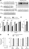
Effects of epoxomicin on the UPS proteins and synaptophysin in spinal cord tissues from sham-operated and SNL animals. A–C, E, Rats with SNL or sham surgery received vehicle or epoxomicin (0.6 nmol/dose) twice daily for 7 d and ipsilateral quadrants of the spinal cord relative to the injury side were taken for analysis 18 h (A, B, C) or 23 h (E) after the last injection, or 1 h after the first daily injection (E). A, Representative Western blot images. c, Sham-vehicle; ce, sham-epoxomicin; s, SNL-vehicle; se, SNL-epoxomicin. B, Levels of E1A ubiquitin-activating enzyme, 20S proteasome subunit, free ubiquitin, ubiquitilated proteins, and ubiquitin C-terminal hydrolase L1. Error bars indicate mean ± SEM; the levels in sham-operated animals are taken as a unit. *p ≤ 0.05; **p ≤ 0.01; ***p ≤ 0.001; n = 4–10. The key in B also applies to C. C, Levels of synaptophysin. D, Inhibition of chymotrypsin-like proteasome activity in MIN6 cells incubated with 20 nm epoxomicin for 18 h. Data are presented as mean ± SEM; n = 2 for the control group, n = 3 for the epoxomicin-treated group. E, Chymotrypsin-like activity in spinal cord tissues from control and SNL animals. Animals were killed before the first daily injection (open bars; n = 4–6) or 1 h after the first daily injection (gray bars; n = 8–10). Clasto-lactacystin-β-lactone (L-β-L) was added to the pooled samples from first experiment to test whether hydrolysis of fluorogenic substrate Suc-LLVY-AMC was catalyzed by the proteasome. Data are shown as mean ± SEM.
Figure 6.
Figure 6.
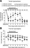
Epoxomicin abolished dynorphin-induced tactile allodynia and thermal hyperalgesia. A, B, Responsive thresholds to innocuous mechanical (von Frey filaments; A) and thermal (52°C hot-plate; B) stimuli. Dynorphin A (15 nmol) was injected intrathecally and twice-daily injections of epoxomicin or vehicle were initiated at the same time. Rats that received dynorphin and vehicle developed enhanced responses to tactile and thermal stimuli, whereas those treated with epoxomicin did not. Termination of epoxomicin resulted in a decreased response thresholds to tactile and thermal stimuli. *p ≤ 0.05, compared to the predynorphin or vehicle baseline values within each treatment group. Data are shown as mean ± SEM; n = 4–6 per group.
Figure 7.
Figure 7.
The proteasome inhibitor epoxomicin prevents morphine-induced tactile allodynia and thermal hyperalgesia. Rats were prepared with either placebo or morphine (2 × 75 mg) pellets implanted subcutaneously. The rats also received vehicle or epoxomicin (0.6 nmol, i.t.) twice daily. A, B, Morphine-treated rats receiving vehicle intrathecally developed tactile allodynia (A) and thermal hyperalgesia (B), as indicated by the reductions in paw withdrawal thresholds and hot-plate latencies. In contrast, morphine-treated rats receiving epoxomicin did not develop these reductions in behavioral endpoints. Data are shown as mean ± SEM. n = 4–7 per group.

Similar articles

Cited by

References

    1. Adams J, Behnke M, Chen S, Cruickshank AA, Dick LR, Grenier L, Klunder JM, Ma YT, Plamondon L, Stein RL. Potent and selective inhibitors of the proteasome: dipeptidyl boronic acids. Bioorg Med Chem Lett. 1998;8:333–338. - PubMed
    1. An JY, Seo JW, Tasaki T, Lee MJ, Varshavsky A, Kwon YT. Impaired neurogenesis and cardiovascular development in mice lacking the E3 ubiquitin ligases UBR1 and UBR2 of the N-end rule pathway. Proc Natl Acad Sci USA. 2006;103:6212–6217. - PMC - PubMed
    1. Angst MS, Clark JD. Opioid-induced hyperalgesia: a qualitative systematic review. Anesthesiology. 2006;104:570–587. - PubMed
    1. Bachar O, Adner M, Uddman R, Cardell LO. Toll-like receptor stimulation induces airway hyper-responsiveness to bradykinin, an effect mediated by JNK and NF-kappa B signaling pathways. Eur J Immunol. 2004;34:1196–1207. - PubMed
    1. Bakalkin G, Yakovleva T, Terenius L. NF-kappa B-like factors in the murine brain. Developmentally regulated and tissue-specific expression. Brain Res Mol Brain Res. 1993;20:137–146. - PubMed

Publication types

MeSH terms